At WiseGEEK, we're committed to delivering accurate, trustworthy information. Our expert-authored content is rigorously fact-checked and sourced from credible authorities. Discover how we uphold the highest standards in providing you with reliable knowledge.
What is the Medial Pterygoid?
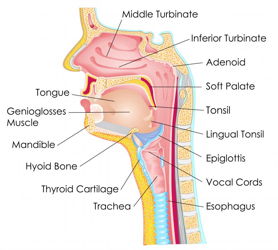
The medial pterygoid is a muscle of the face, found inside of the jawbone. It is a nearly vertical muscle, its fibers running from the inner surface of the upper jaw, just behind the top teeth, to the angle of the mandible, which is the curve at the corner of the jawbone. Also known as the internal pterygoid or pterygoidus internus, it is used for chewing. By elevating the mandible, it closes the jaw. It also has a minor involvement in side-to-side jaw movements.
Stretching almost perpendicular to the other pterygoid muscle, the pterygoidus externus, the medial pterygoid has two separate heads or sections. The deep head, which consists of the majority of the muscle’s fibers, originates on the inner part of a bony surface known as the lateral pterygoid plate. Found on the sphenoid bone, a butterfly-shaped bone on the inside of the face, the lateral pterygoid plate is the surface of that bone that protrudes the farthest toward either side of the face and is situated behind the top row of teeth and in front of the ear. The deep head of the medial pterygoid arises from the internal surface of this plate.

This muscle’s superficial head, the one nearer to the skin, accounts for a much smaller portion of the internal pterygoid’s surface area. It arises from a surface on the maxilla, the bone of the upper jaw, known as the maxillary tuberosity, which is a rounded bony prominence found on either side of the maxilla near the upper wisdom teeth. The superficial head also originates on a small area of the back outer corner of the palatine bone, which is the flat bone that forms the roof of the mouth.
From these points of origin, the fibers of the medial pterygoid form a muscle that is quadrilateral in shape as it crosses the jaw. This muscle, angling slightly outward and backward as it descends, attaches along the inner surface of the angle and ramus of the mandible. These are, respectively, the curve at the outer corner of the jawbone and the ascending portion of the outer jawbone that angles toward the earlobe. At these insertion points, the fibers of the medial pterygoid unite with those of the masseter muscle, the main chewing muscle of the jaw. This common point of insertion permits the pterygoid to assist the masseter in lifting the mandible during chewing and other closing movements.
AS FEATURED ON:
AS FEATURED ON:

-
![The mandible is the lower part of the jaw, which opens and closes like a hinge thanks to a network of muscles.]() By: snapgalleriaThe mandible is the lower part of the jaw, which opens and closes like a hinge thanks to a network of muscles.
By: snapgalleriaThe mandible is the lower part of the jaw, which opens and closes like a hinge thanks to a network of muscles.
Discuss this Article
Post your comments