At WiseGEEK, we're committed to delivering accurate, trustworthy information. Our expert-authored content is rigorously fact-checked and sourced from credible authorities. Discover how we uphold the highest standards in providing you with reliable knowledge.
What is the Anatomy of the Bladder?
The anatomy of the bladder is designed so it is capable of holding enough urine that an individual only needs to relieve him or herself four to six times each day. It is located in the pelvic cavity, near the small intestine and the reproductive organs. About the size and shape of a lemon, the surface of the bladder allows the organ to stretch as it fills.
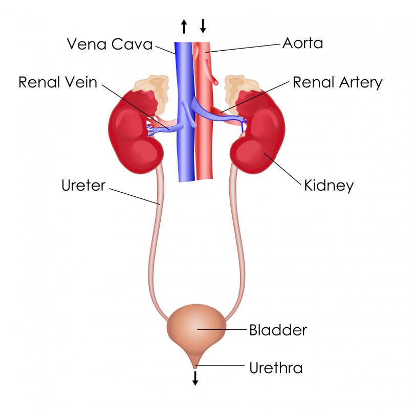
The bladder is connected to the kidneys by tubes called ureters, which join the bladder at the ureter orifices. These tubes enter the bladder through the top on the left and right sides, and pass the urine into the bladder, where it is stored until elimination. During the process of elimination, the urine passes out of the bladder through a structure in the bottom, called the urethra. At the central point between these three openings is a pale, triangular region known as the trigonum vesicae, which is used primarily by medical professionals as a reference point for other parts of the anatomy of the bladder.

Several layers of tissue and muscle make up the surface portion of the anatomy of the bladder. The outermost layer is called the serosa, or peritoneum. This thin tissue is similar to the tissues that line the surfaces of other organs in the abdomen, such as the kidneys and the inside of the abdomen itself. Underneath this thin layer of tissue is a layer that includes the muscles that involuntarily control the expansion and contraction of the bladder. This layer, called the detrusor muscle, works involuntarily to expand as urine drains in from the ureters, and to contract as it is eliminated from the body.

On the inside of the bladder is a mucous membrane that has elastic qualities and forms folds when the bladder is empty. It is attached to the outer muscle layer by a structure called the sub-mucous layer. This layer shares the same elasticity as other layers of the bladder.
The muscle that holds the urine in the bladder is known as the internal urethral sphincter. It is this valve that opens during the process of elimination in order to allow the urine to pass through. In some individuals, incontinence is caused by a malfunction of this valve.

The anatomy of the bladder is a simple structure that works on a largely involuntary basis to allow an individual to wait hours between eliminating urine from the body. The basic anatomy of the bladder is the same in men and women, with the length of the urethra the main difference. The size and shape of the bladder can change as an individual ages, and may hold less the older the person gets.
AS FEATURED ON:
AS FEATURED ON:

-
![A cutaway of a female body showing the bladder in dark pink.]() By: kocakayaaliA cutaway of a female body showing the bladder in dark pink.
By: kocakayaaliA cutaway of a female body showing the bladder in dark pink. -
![The bladder is part of the larger urinary system, which filters out waste from the body.]() By: snapgalleriaThe bladder is part of the larger urinary system, which filters out waste from the body.
By: snapgalleriaThe bladder is part of the larger urinary system, which filters out waste from the body. -
![A urologist is a medical doctor who specializes in diseases and conditions of the urinary system.]() By: Darren BakerA urologist is a medical doctor who specializes in diseases and conditions of the urinary system.
By: Darren BakerA urologist is a medical doctor who specializes in diseases and conditions of the urinary system.


Discuss this Article
Post your comments