At TheHealthBoard, we're committed to delivering accurate, trustworthy information. Our expert-authored content is rigorously fact-checked and sourced from credible authorities. Discover how we uphold the highest standards in providing you with reliable knowledge.
What is Superficial Spreading Melanoma?
Superficial spreading melanoma (SSM) is a type of skin lesion which may be a precursor to skin cancer. It initially appears small and looks similar to a slightly large freckle, but significantly progresses into a larger, more prominent lesion. Anyone may contract this condition regardless of age and gender, though certain people are more susceptible than others. While its prognosis is generally considered promising and not life-threatening, the condition may be fatal if left untreated.
Of the four types of melanoma, superficial spreading melanoma accounts for about 70 percent of all reported cases. Nodular melanoma accounts for about 15 percent of cases, lentigo maligna melanoma for about 10 percent, and acral lentiginous melanoma for about 5 percent. The latter of these is considered the most life-threatening of all forms of melanoma. Since superficial spreading melanoma is quite prominent, it is still the leading cause of death from cancer despite its positive prognosis.

The condition is most commonly found among females, though men may contract it as well. Females will usually find lesions on their legs, while men will generally find lesions scattered among the upper torso, particularly between the neck and pelvis. Superficial spreading melanoma usually occurs post-puberty and tends to be more prevalent among Caucasians than people of other ethnic backgrounds. It is additionally more common among people who have 100 or more moles on their bodies.

Two stages of superficial spreading melanoma denote both the appearance of the condition and the threat posed to its carrier. The initial phase is known as the radial phase, in which lesions first form on the skin. These lesions appear small and thin, and may remain in this phase for a few months or even years. The radial phase poses the least significant threat of cancer.

The second stage of the condition, called the vertical growth phase, proves significantly more life-threatening. Lesions grow in excess of about .25 inches (6 mm) and move deeper into the surface of the skin, possibly causing pain, skin irritation, oozing, or bleeding. The melanoma may spread to surrounding skin tissue, and its pigmentation may darken. A slight diminishing of this pigmentation sometimes occurs as the immune system attempts to fight the condition, but the melanoma still exists and requires treatment.

Many different factors contribute to the likelihood of contracting superficial spreading melanoma. Patients often have a sibling or parent with the condition. Other uncontrollable factors, such as a weak immune system or a rare hereditary skin disease called xeroderma pigmentosum, may pose further risk. Sun exposure, a more manageable factor, adds to the likelihood of superficial spreading melanoma, particularly during adolescence.

It is recommended that a dermatologist is consulted at any possible signs of this condition. Superficial spreading melanoma is generally diagnosed via biopsy, though sometimes x-rays, computerized axial tomography (CT) scans, magnetic resonance imaging (MRIs), ultrasounds, and other procedures are employed. The condition is commonly treated through surgical removal, which significantly reduces the risk of its development into potentially fatal skin cancer.
AS FEATURED ON:
AS FEATURED ON:

-
![Excessive UV exposure can lead to skin cancer, a serious disease of the integumentary system.]() By: Amy WaltersExcessive UV exposure can lead to skin cancer, a serious disease of the integumentary system.
By: Amy WaltersExcessive UV exposure can lead to skin cancer, a serious disease of the integumentary system. -
![Most cases of superficial spreading melanoma stem from over-exposure to the sun, particularly during adolescence.]() By: AntonioguillemMost cases of superficial spreading melanoma stem from over-exposure to the sun, particularly during adolescence.
By: AntonioguillemMost cases of superficial spreading melanoma stem from over-exposure to the sun, particularly during adolescence. -
![The prognosis of superficial spreading melanoma is generally considered promising.]() By: Lisa F. YoungThe prognosis of superficial spreading melanoma is generally considered promising.
By: Lisa F. YoungThe prognosis of superficial spreading melanoma is generally considered promising. -
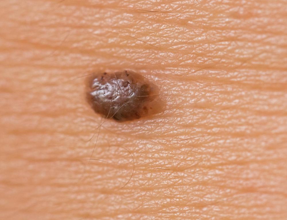
![Moles may be malignant if they are asymmetrical, have irregular borders, appear to spread their color into surrounding skin or change in appearance.]() By: bertys30Moles may be malignant if they are asymmetrical, have irregular borders, appear to spread their color into surrounding skin or change in appearance.
By: bertys30Moles may be malignant if they are asymmetrical, have irregular borders, appear to spread their color into surrounding skin or change in appearance. -
![The appearance of moles might be a sign of melanoma.]() By: F.C.G.The appearance of moles might be a sign of melanoma.
By: F.C.G.The appearance of moles might be a sign of melanoma. -
![Melanomas are commonly found on areas of the body that are frequently exposed to sunlight.]() By: LoloStockMelanomas are commonly found on areas of the body that are frequently exposed to sunlight.
By: LoloStockMelanomas are commonly found on areas of the body that are frequently exposed to sunlight. -
![In many cases, moles should be checked under a microscope after removal to ensure they are benign.]() By: luchschen_shutterIn many cases, moles should be checked under a microscope after removal to ensure they are benign.
By: luchschen_shutterIn many cases, moles should be checked under a microscope after removal to ensure they are benign.


Discuss this Article
Post your comments