At WiseGEEK, we're committed to delivering accurate, trustworthy information. Our expert-authored content is rigorously fact-checked and sourced from credible authorities. Discover how we uphold the highest standards in providing you with reliable knowledge.
What is an Electroretinography?
 Mary McMahon
Mary McMahon
Mary McMahon
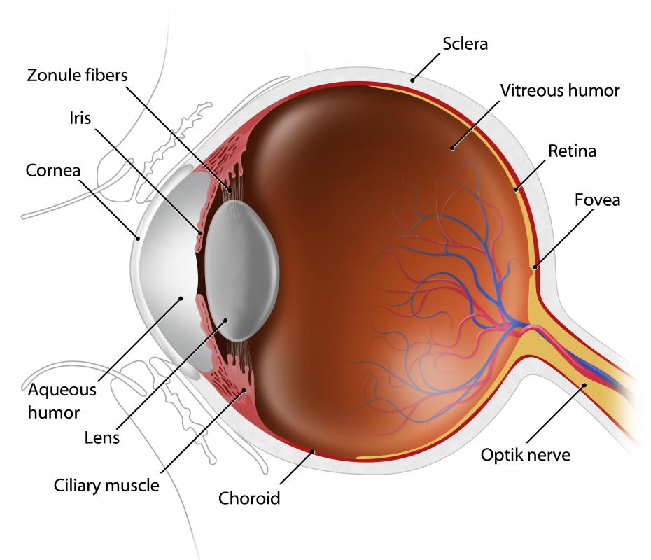
Electroretinography (ERG) is a procedure in which the sensitivity of the retina to light is measured by recording electrical responses generated inside the eye when it is exposed to stimuli. This test is usually ordered when a doctor suspects that a patient has a retinal disorder, whether acquired or congenital. It is relatively low risk when performed by an experienced technician, and can even be performed on children and infants, although sedation may be required for younger patients so that they do not move during the testing.
In an electroretinography test, the patient is usually asked to lie down. The pupils are dilated with eyedrops, and a numbing agent is applied to the eye so that an electrode can be gently placed on the cornea while another electrode is placed near the eye. To prevent the patient from blinking, the eye is held open with a speculum and a lubricant is applied to minimize discomfort. Then, the patient is exposed to a set of standardized stimuli, and the electrical responses are recorded.

For the second part of the test, the room is darkened and the patient's eyes are allowed to adjust so that the test can be repeated. This part of the electroretinography screening tests the rods, the light sensitive components in the retina, as opposed to the cones, the structures in the retina which are sensitized to color. Once the test is complete, the electrodes can be removed and the patient can be allowed to sit up again.

The results are available instantly, in the form of the readout generated by the electrodes. Electroretinography can reveal disorders which span the entire retina, and provide more information about retinal function. However, this test is not suited to looking for things like macular degeneration, which involve only a small area of the retina. Additional screening may be needed after an electroretinogram has been generated to learn more about what is going on inside the patient's eyes.

Patients should prepare for a test which will take around an hour from start to finish. Because the eyes are still dilated at the end of the exam, it is strongly recommended to secure a ride home with a friend or family member, as it can be dangerous to drive and challenging to navigate public transit when the eyes are highly sensitive to light. Patients should also avoid touching their eyes for at least an hour after the exam, and they should report any complications of the electroretinography such as pain or scratches on the cornea to their physicians.
Mary McMahon
Ever since she began contributing to the site several years ago, Mary has embraced the exciting challenge of being a WiseGEEK researcher and writer. Mary has a liberal arts degree from Goddard College and spends her free time reading, cooking, and exploring the great outdoors.
Learn more...
Mary McMahon
Ever since she began contributing to the site several years ago, Mary has embraced the exciting challenge of being a WiseGEEK researcher and writer. Mary has a liberal arts degree from Goddard College and spends her free time reading, cooking, and exploring the great outdoors.
Learn more...AS FEATURED ON:
AS FEATURED ON:

-
![In an electroretinography test, the patient's pupils are dilated with eyedrops.]() By: ra2 studioIn an electroretinography test, the patient's pupils are dilated with eyedrops.
By: ra2 studioIn an electroretinography test, the patient's pupils are dilated with eyedrops. -
![Patients should not drive immediately after having an electroretinography.]() By: Subbotina AnnaPatients should not drive immediately after having an electroretinography.
By: Subbotina AnnaPatients should not drive immediately after having an electroretinography. -
![During electroretinography, an electrode is placed on the patient's cornea.]() By: kocakayaaliDuring electroretinography, an electrode is placed on the patient's cornea.
By: kocakayaaliDuring electroretinography, an electrode is placed on the patient's cornea.


Discuss this Article
Post your comments