At WiseGEEK, we're committed to delivering accurate, trustworthy information. Our expert-authored content is rigorously fact-checked and sourced from credible authorities. Discover how we uphold the highest standards in providing you with reliable knowledge.
What are the Best Tips for EEG Analysis?
An electroencephalogram (EEG) is a process by which researchers and neurologists record the electrical activity of the nerve cells in the brain by several electrodes attached to the scalp. After 20 to 40 minutes of data collection, physicians can assess the pattern of oscillations coming from the synchronized activity of large groups of nerve cells for abnormalities that reflect brain dysfunction or disease. The most commonly observed wave patterns, including the alpha, beta, and delta waves, fluctuate in the one-to-20-megahertz (MHz) frequency range, with each wave having its own frequency sub range. Through EEG analysis, neurologists can identify abnormal wave patterns and localize anomalous brain activity.
EEGs demonstrate alpha waves, ranging from eight to 13 MHz, emanating from the posterior parts of the brain, with higher amplitude waves on the dominant side of the brain. Alpha waves occur when the patient is relaxed with the eyes closed. The waves decrease in height when the eyes are open. Abnormal alpha activity on EEG analysis may indicate a comatose condition. A difference in magnitude of the waves between the two sides that exceeds 50 percent indicates posterior brain abnormalities.

Delta waves, with slow frequencies of one to four MHz, can be recorded in the frontal areas of most adults. These waves occur with normal sleep, but they may be abnormally present in patients with hydrocephalus (too much fluid on the brain), metabolic imbalances, and deep brain tissue lesions. On the other hand, the beta waves, typically at frequencies greater than 13 MHz, are also found frontally in patients who are alert, active, and busy. Upon EEG analysis, beta waves may be blunted in patients who have taken sedatives.

Mu waves occur in the same frequency as the alpha waves, and they originate from the areas of the brain that control the senses and movement. The waves occur more prominently during states of relaxation than during periods of alert activity. In cases where excessive mu waves occur, autism may be a possible diagnosis. Theta waves occur with drowsiness or arousal, and they are measured in areas apart from where the mind is actively involved in a task. Theta waves are noted in EEG analysis when the patient is attempting to suppress a thought or avoid an action.

Spikes and strong wave fluctuations may reflect seizure activity in cases of epilepsy, drug-induced seizures, or trauma-related convulsions. During EEG analysis, these rapid, transient wave alterations must be interpreted with a view toward the potential for artifacts produced by the environment, muscle activity, ocular fluttering or movement, and tongue movement. Artifact is a critical consideration when an EEG occurs in a patient with Parkinson’s disease or tremor. Artifacts from the heartbeat can also produce “noise” on an EEG reading.
AS FEATURED ON:
AS FEATURED ON:

-
![An electroencephalogram (EEG) is a process to record the electrical activity of the nerve cells in the brain using several electrodes that are attached to the scalp.]() By: TobilanderAn electroencephalogram (EEG) is a process to record the electrical activity of the nerve cells in the brain using several electrodes that are attached to the scalp.
By: TobilanderAn electroencephalogram (EEG) is a process to record the electrical activity of the nerve cells in the brain using several electrodes that are attached to the scalp. -
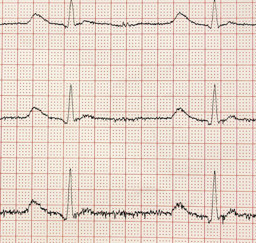
![Spikes and strong wave fluxuations noted during an EEG analysis may reflect seizure activity in cases of epilepsy.]() By: a40757seSpikes and strong wave fluxuations noted during an EEG analysis may reflect seizure activity in cases of epilepsy.
By: a40757seSpikes and strong wave fluxuations noted during an EEG analysis may reflect seizure activity in cases of epilepsy. -
![A doctor may order an EEG for patients suffering from headaches.]() By: JPC-PRODA doctor may order an EEG for patients suffering from headaches.
By: JPC-PRODA doctor may order an EEG for patients suffering from headaches. -
![EEG analysis can identify abnormal wave patterns and irregular brain activity.]() By: NaeblysEEG analysis can identify abnormal wave patterns and irregular brain activity.
By: NaeblysEEG analysis can identify abnormal wave patterns and irregular brain activity.


Discuss this Article
Post your comments