At WiseGEEK, we're committed to delivering accurate, trustworthy information. Our expert-authored content is rigorously fact-checked and sourced from credible authorities. Discover how we uphold the highest standards in providing you with reliable knowledge.
What is the Optic Disc?
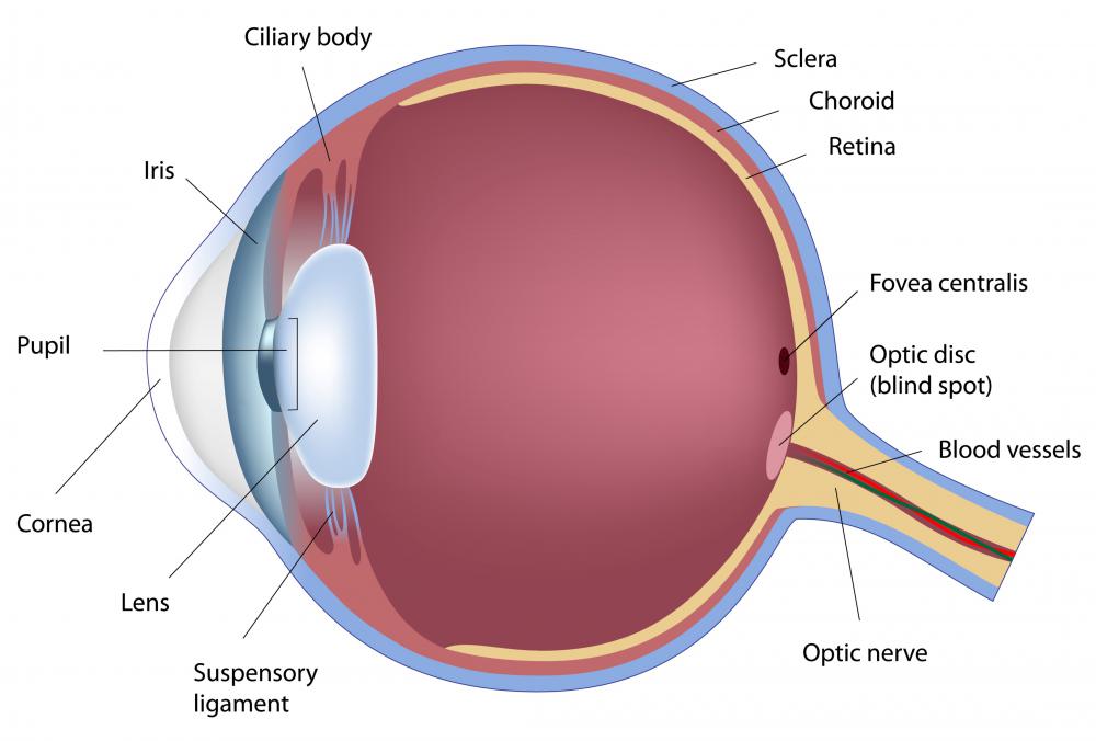
The optic disc is a vertically oval spot in the back of the eye about three to four millimeters (0.14 to 0.18 inch) nasal to the center. It is made up of the nerve fibers of nerve cells, called ganglion cells, that reside within the light-sensitive layer of the back of the eye, called the retina. The 1.0 to 1.2 million ganglion cell nerve fibers or axons leave the eye at the disc and form the optic nerve, which carries visual information to the brain. A physiological blind spot in the visual field of each eye results from the lack of light-sensitive cells, rods and cones, at the optic disc. An examination of the optic disc through the pupil yields valuable diagnostic information regarding various ocular and neurological diseases.
On average, the optic disc measures approximately 1.92 millimeters (0.09 inch) vertically by 1.76 millimeters (0.08 inch) horizontally. The optic cup is a small central depression in the disc, usually spanning about 33 percent of the disc area. Normally, the disc is orange to yellowish-pink in color with well-defined margins. The rim around the cup is slightly thicker at the inferior pole, with the thinnest portion of the border located temporally. A standard ophthalmic examination includes notation of the disc color, cup size, margin definition, associated bleeding or swelling, and rim anomalies.

Glaucoma is a degenerative optic nerve disease typically associated with a sustained elevation of the eye pressure. A characteristic feature of glaucoma is progressive expansion of the optic cup relative to the size of the optic disc. Notching of the disc rim, as well as bleeding at the disc margin, may also occur. Progressive optic disc cupping is a sign of continuing retinal nerve fiber layer thinning. Clinical studies show that lowering the eye pressure by 20 to 30 percent effectively halts the optic nerve damage in most cases.

A pale disc is indicative of poor blood supply or atrophy. Optic atrophy is the hallmark of ganglion cell damage. Severe degrees of damage are characterized by a chalky-white disc color with stark, unusually sharp borders. Mild degrees of atrophy may be recognized by comparison of the disc color with the fellow eye. Optic atrophy occurs four to six weeks after cell damage due to reduced blood circulation or inflammation.

Optic disc swelling or edema occurs due to impaired flow of nutrients through the axons. This can result from increased pressure in the head, reduced blood flow, inflammation, or mechanical compression. Optic nerve edema features include blurring of the disc margins, bleeding around the disc, elevation of the nerve head, and a reddish color of the disc. Disc swelling may be a sign of a brain tumor, an orbital tumor, active optic nerve inflammation, or a mini-stroke to the nerve.

Optic disc drusen are calcific nodules buried with the optic nerve head. Drusen cause an elevation of the optic nerve head with a scalloped appearance. They are bilateral in 75 percent to 86 percent of cases. Although drusen usually produce no symptoms, transient visual fluctuations and minor visual field defects are sometimes reported.
AS FEATURED ON:
AS FEATURED ON:

-
![The optic disc lies in the back of the eye.]() By: Alila Medical MediaThe optic disc lies in the back of the eye.
By: Alila Medical MediaThe optic disc lies in the back of the eye. -
![The optic disc of the eye is made up of nerve fibers and nerve cells.]() By: Péter MácsThe optic disc of the eye is made up of nerve fibers and nerve cells.
By: Péter MácsThe optic disc of the eye is made up of nerve fibers and nerve cells. -
![Glaucoma causes a progressive expansion of the optic cup relative to the size of the optic disc.]() By: joshyaGlaucoma causes a progressive expansion of the optic cup relative to the size of the optic disc.
By: joshyaGlaucoma causes a progressive expansion of the optic cup relative to the size of the optic disc. -
![Glaucoma is a degenerative optic nerve disease that is typically associated with an sustained elevation of the eye pressure.]() By: JackFGlaucoma is a degenerative optic nerve disease that is typically associated with an sustained elevation of the eye pressure.
By: JackFGlaucoma is a degenerative optic nerve disease that is typically associated with an sustained elevation of the eye pressure. -
![Optic disc swelling may be a sign of a brain tumor.]() By: CLIPAREA.comOptic disc swelling may be a sign of a brain tumor.
By: CLIPAREA.comOptic disc swelling may be a sign of a brain tumor. -
![Individuals suffering from glaucoma may experience tunnel vision.]() By: JPC-PRODIndividuals suffering from glaucoma may experience tunnel vision.
By: JPC-PRODIndividuals suffering from glaucoma may experience tunnel vision. -
![Optic disc swelling occurs due to impaired flow of nutrients through the axons.]() By: Photographee.euOptic disc swelling occurs due to impaired flow of nutrients through the axons.
By: Photographee.euOptic disc swelling occurs due to impaired flow of nutrients through the axons. -
![A person suffering from optic disc swelling may experience frequent headaches.]() By: Syda ProductionsA person suffering from optic disc swelling may experience frequent headaches.
By: Syda ProductionsA person suffering from optic disc swelling may experience frequent headaches.


Discuss this Article
Post your comments