At WiseGEEK, we're committed to delivering accurate, trustworthy information. Our expert-authored content is rigorously fact-checked and sourced from credible authorities. Discover how we uphold the highest standards in providing you with reliable knowledge.
What Is the Endolymphatic Sac?
The endolymphatic sac is a structure at the end of the membranous labyrinth in the inner ear. Endolymph is the name of the fluid which fills the membranous labyrinth. The endolymphatic sac has an irregular, complex shape and is made up of interconnected tubes and spaces. Its function is thought to involve absorbing and secreting as well as playing a part in the immune response. Although its role is not fully understood, the endolymphatic sac is thought to be involved in the development of Meniere's disease, a condition in which dizziness and hearing problems are experienced.
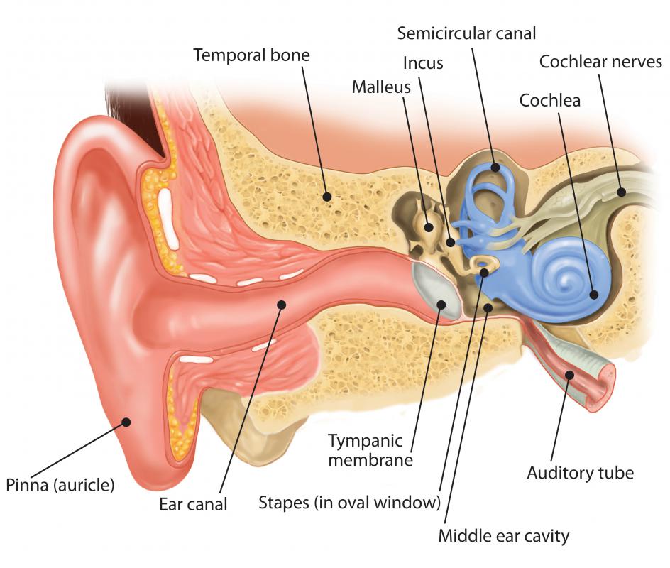
Inside the bony labyrinth of the inner ear, which is filled with a fluid known as perilymph, lies the membranous labyrinth. The membranous labyrinth is completely sealed so that the endolymph inside it does not communicate with the perilymph outside. Included in the membranous labyrinth are the sensory structures involved with balance and hearing. Those concerned with balance are the semicircular canals, saccule and utricle, while the cochlea is the structure involved with hearing. These sensory parts of the labyrinth are connected to two non-sensory structures, which are the ductus endolymphaticus and the endolymphatic sac.

Endolymph from the cochlea and the saccule passes through the tube, or duct, known as the ductus endolymphaticus. From the semicircular canals and the utricle, endolymph passes through the ductus utriculosaccularis, which joins the ductus endolymphaticus. The ductus endolymphaticus leads through a channel called the aquaeductus vestibuli to the endolymphatic sac. This sits inside the temporal bone in the skull, forming the blind ending of the membranous labyrinth. Although all the sections of the membranous labyrinth are connected by ducts, endolymph does not actually flow through the compartments.
The endolymphatic sac has a simple structure in newborns, but after the first year of life it begins to develop a more complex shape. At about the age of four it will have developed the complicated structure which is found in adults. The middle section of the sac is the most complex and is known as the tubular portion. Only a small amount of endolymph is found inside the sac and, unlike most of the membranous labyrinth, it is not surrounded by perilymph.
Although researchers continue to investigate the processes involved in Meniere's disease, the endolymphatic sac is thought to play an important part. In patients with the disease, the sac is often small or invisible when viewed using magnetic resonance imaging (MRI) scanning. The use of endolymphatic sac surgery as a treatment for Meniere's disease is controversial, and many doctors consider it ineffective.
AS FEATURED ON:
AS FEATURED ON:

-
![Endolymph from the cochlea and the saccule passes through the tube, or duct, known as the ductus endolymphaticus.]() By: kocakayaaliEndolymph from the cochlea and the saccule passes through the tube, or duct, known as the ductus endolymphaticus.
By: kocakayaaliEndolymph from the cochlea and the saccule passes through the tube, or duct, known as the ductus endolymphaticus.
Discuss this Article
Post your comments