At WiseGEEK, we're committed to delivering accurate, trustworthy information. Our expert-authored content is rigorously fact-checked and sourced from credible authorities. Discover how we uphold the highest standards in providing you with reliable knowledge.
What is Gestational Trophoblastic Disease?
Gestational trophoblastic disease (GTD) is the term for uterine tumors that develop from the trophoblast – a layer of cells that surrounds an embryo during pregnancy. These cells normally develop into the placenta, which delivers nutrients to a fetus; in rare instances, though, they can develop into tumors instead. There are four primary types of gestational trophoblastic disease: hydatidiform mole, invasive mole, choriocarcinoma, and placental site trophoblastic tumor.
Hydatidiform mole GTD, or molar pregnancy, can occur when one or more sperm cells fertilize an egg that has no nucleus or deoxyribonucleic acid (DNA). It can also be caused by two sperm fertilizing an egg that does contain DNA. The first type of fertilization produces no fetal tissue at all, and the second type may produce fetal tissue but not a viable fetus.
Invasive mole is the type of GTD that develops when hydatidiform moles penetrate the uterine muscle layer. This type of gestational trophoblastic disease can occur in women who have had hydatidiform moles removed. It is more common in women over 40.
Choriocarcinoma GTD is malignant and tends to spread quickly. This type of GTD often spreads beyond the uterus. It can develop from a hydatidiform mole, or it can occur after a full-term pregnancy or a pregnancy that was terminated early.

Placental site trophoblastic tumor is the rarest type of GTD. It occurs when the placenta becomes attached to the uterus, and usually occurs after pregnancy. These types of tumors usually do not spread beyond the uterus and are typically unresponsive to chemotherapy.
There are some times when gestational trophoblastic disease occurs during an otherwise viable pregnancy. In these instances, it is quite rare for an embryo or fetus to survive. Such pregnancies usually end in miscarriage or fetal death.
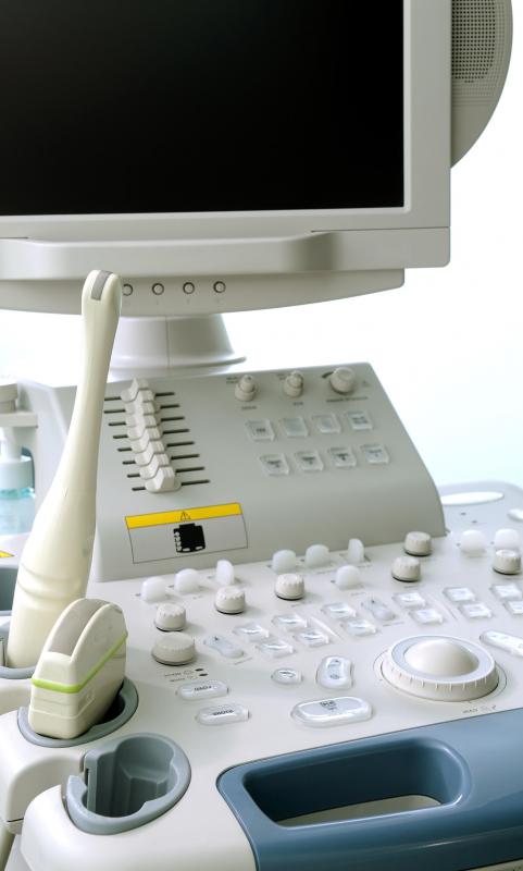
Gestational trophoblastic disease symptoms include an abnormally large uterus, excessive vomiting, vaginal bleeding, and preeclampsia. The disease is usually found during a routine pregnancy ultrasound. It can also be diagnosed by testing for high levels of human chorionic gonadotropin (HCG) in the blood. Diagnosis is usually confirmed with a biopsy.
GTD is typically treatable through surgery, chemotherapy, or radiation. Tumors can often be suctioned out of the uterus. Most women who have GTD once are able to have subsequent successful pregnancies.
Risk factors for GTD include becoming pregnant before the age of 20 or after the age of 40, a history of miscarriage, and a history of difficulty becoming pregnant. Family history does not appear to play a significant role, though there have been reported instances of many women in a family having GTD. Also, gestational trophoblastic disease is slightly more common in women with blood type A or AB than in women with blood type B or O.
AS FEATURED ON:
AS FEATURED ON:

-
![Gestational trophoblastic disease can be detected during ultrasound.]() By: Andrey ChmelyovGestational trophoblastic disease can be detected during ultrasound.
By: Andrey ChmelyovGestational trophoblastic disease can be detected during ultrasound.
Discuss this Article
Post your comments