At TheHealthBoard, we're committed to delivering accurate, trustworthy information. Our expert-authored content is rigorously fact-checked and sourced from credible authorities. Discover how we uphold the highest standards in providing you with reliable knowledge.
What is Echocardiography?
 Mary McMahon
Mary McMahon
Mary McMahon
Echocardiography is a type of medical imaging which is used to visualize the heart for the purpose of evaluating heart conditions or diagnosing suspected heart problems. It is also known as cardiac ultrasound, and may be referred to as a “cardiac echo” or “echo” for short. Depending on the type of echocardiography being performed, this type of ultrasound imaging can be very invasive or minimally invasive, and it is typically performed as an outpatient procedure in a hospital or clinic.
In echocardiography, the technician uses a transducer which emits high frequency sound waves to generate a picture of the heart. The transducer reads the sound waves when they return, creating a map of the inside of the chest on the basis of the ways in which the sound waves change. Modern ultrasound machines are capable of generating very high resolution images, and it is also possible to create a three dimensional echocardiogram which provides an even higher level of detail.

In basic echocardiography, the transducer is manipulated along the chest, with a conductive gel ensuring that the sound waves are transmitted through the chest wall. In a transesophageal echo, the ultrasound transducer is actually inserted into the throat, providing a clear image without interference from the ribs. A basic echocardiography procedure is non-invasive and comfortable for the patient, while a transesophageal procedure can be extremely uncomfortable, although it provides a more reliable image.

A variety of types of images of the heart can be acquired with cardiac ultrasound. Still images providing information about the structures of the heart can be very useful, as can video images which document the heart in action. Video images can be used to measure the output of the heart, and it is also possible to follow dyes and tracers to check for leaks around the valves. Some ultrasound machines generate colored images for greater contrast to make the echocardiogram easier to read.

This procedure can be performed by a doctor or by an ultrasound technician. If the test is performed by a technician, the patient may need to wait several hours or days for the test results to be interpreted by a radiologist or cardiac specialist, while a doctor may be able to discuss the results right away. Echocardiography is only one tool in an arsenal of options which can be used to study the heart for medical reasons, but it can provide a great deal of information with a minimum of discomfort and expense for the patient.
Mary McMahon
Ever since she began contributing to the site several years ago, Mary has embraced the exciting challenge of being a TheHealthBoard researcher and writer. Mary has a liberal arts degree from Goddard College and spends her free time reading, cooking, and exploring the great outdoors.
Learn more...
Mary McMahon
Ever since she began contributing to the site several years ago, Mary has embraced the exciting challenge of being a TheHealthBoard researcher and writer. Mary has a liberal arts degree from Goddard College and spends her free time reading, cooking, and exploring the great outdoors.
Learn more...AS FEATURED ON:
AS FEATURED ON:

-
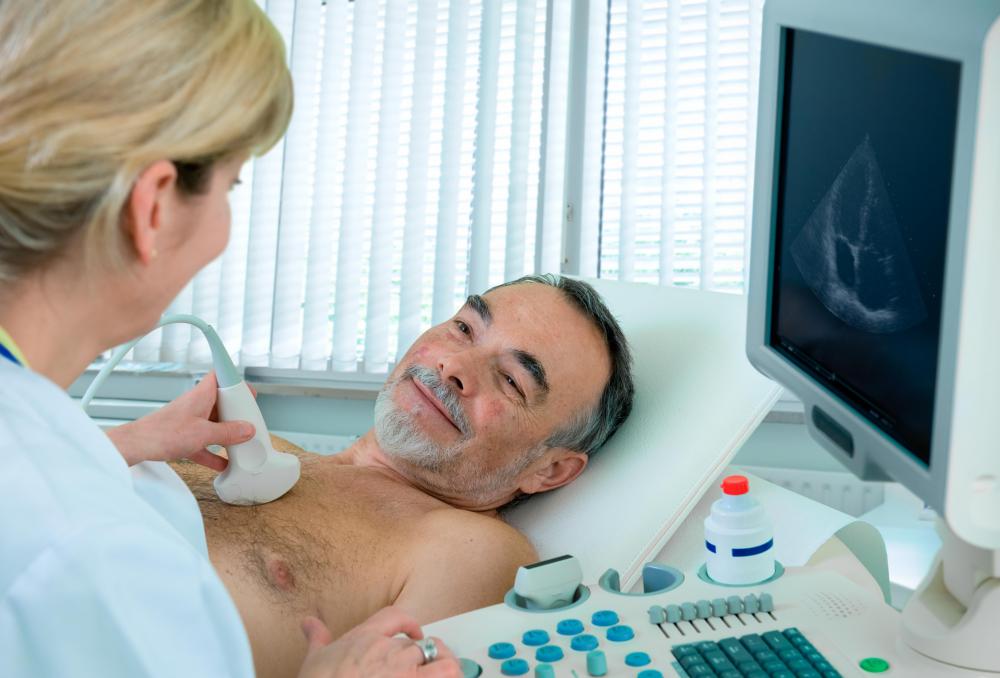
![Echocardiography uses sound waves to monitor the condition of a patient's heart.]() By: Alexander RathsEchocardiography uses sound waves to monitor the condition of a patient's heart.
By: Alexander RathsEchocardiography uses sound waves to monitor the condition of a patient's heart. -
![Echocardiography uses sounds waves to visualize the heart.]() By: adimasEchocardiography uses sounds waves to visualize the heart.
By: adimasEchocardiography uses sounds waves to visualize the heart. -
![A diagnostic ultrasound uses high-frequency sound waves to create an image of the inside of a body.]() By: poco_bwA diagnostic ultrasound uses high-frequency sound waves to create an image of the inside of a body.
By: poco_bwA diagnostic ultrasound uses high-frequency sound waves to create an image of the inside of a body. -
![A doctor may be able to discuss the results of an echocardiogram with a patient immediately.]() By: Photographee.euA doctor may be able to discuss the results of an echocardiogram with a patient immediately.
By: Photographee.euA doctor may be able to discuss the results of an echocardiogram with a patient immediately. -
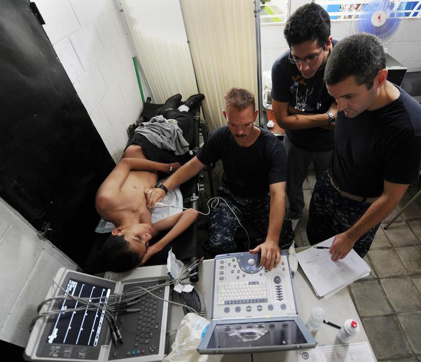
![An echocardiographer employs sonogram technology to provide diagnostic imaging of the heart.]()
An echocardiographer employs sonogram technology to provide diagnostic imaging of the heart.


Discuss this Article
Post your comments