At WiseGEEK, we're committed to delivering accurate, trustworthy information. Our expert-authored content is rigorously fact-checked and sourced from credible authorities. Discover how we uphold the highest standards in providing you with reliable knowledge.
What Is Digital Radiography?
Digital radiography is a type of X-ray imaging in which the images are transposed digitally onto computers or other devices rather than being developed onto film. Instead of using electromagnetic radiation and chemical processing to record an X-ray onto film, digital versions use digital sensors to record images onto an image capture device, which then creates a digital image file. This file can then be used by medical staff members, and the file can be attached to a patient’s medical notes for future reference. It can be printed to paper or slide material so can be used the same as any standard X-ray, but without as much risk and usually at lower overall cost. The initial expenses with a digital system can be immense, but over time they usually pay for themselves. These sorts of imaging devices are most commonly seen in hospitals, specialist medical practices, and dental offices. Dental imaging requires a slightly different process but the technology and basic concept are the same.
Understanding X-Ray Technology Generally

Electromagnetic radiation has been used in the medical profession for imaging and diagnostics for many years. X-radiation technology, known more simply as X-ray technology, allows accurate images to be captured of a person, animal, or thing’s internal composition. A generator uses strong electromagnetic light paired with a detector; most things, humans included, will naturally absorb some of the light, which is what allows the detector to map out images and specific locations.

Most of the earliest X-rays depended on photographic films to capture the images and make them readable. Digital detectors skip this step; rather than using light beamed through objects onto film, it allows for digital scanning and image interpretation. In terms of radiation the two are about the same initially, though digital versions typically have a shorter exposure time and as such tend to be more efficient.
Indirect Conversions

There are usually two types of digital radiography. The first, known as indirect digital radiography, involves amorphous silicon (a-Si) flat panel detectors, and it works by converting X-ray images to light and channeling the image through an amorphous silicon photodiode layer that converts it to a digital signal. Thin film transistors (TFTs) then read this digital output, and it is turned into a data file that can be viewed by the X-ray technician. The technician checks that the X-ray is of a high quality and shows the desired body part clearly, then he or she forwards it electronically to a radiologist for interpretation. This is the most common form and is used for most medical imagery.
Accelerated Imaging

The second type is direct digital radiography and involves amorphous selenium (a-Se) flat panel detectors. This uses a high-voltage electrode to accelerate X-ray photons through a selenium layer, and the pattern is then recorded. This creates an image file that is sent directly to the technician and on to the radiologist.
Special Considerations for Dental Imaging

Digital dental radiography requires a slightly different process. Intraoral images are taken by asking the patient to bite down on an X-ray sensor placed inside the mouth. There is much less radiation involved in digital scans than in film-based electromagnetic radiation, so it is usually safe enough to take numerous X-rays and view all of the teeth from multiple angles. This is useful for checking for decay or tooth problems that can be easily missed during a clinical checkup.

Extraoral images are created by placing the sensor outside the mouth, at the front of the head. This type of image typically shows all of the teeth from the tip to the root, and it is useful for identifying fractures or problems with the mandibular. It is less effective for spotting tooth decay or bone loss.
AS FEATURED ON:
AS FEATURED ON:

-
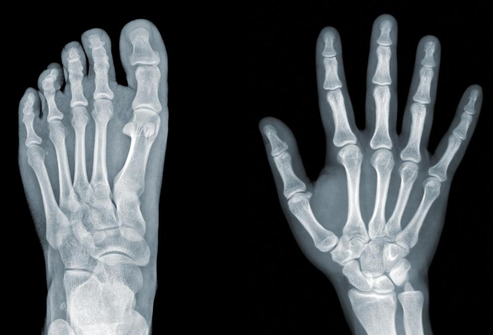
![Digital radiography captures X-ray images on digital equipment as opposed to film.]() By: eAlisaDigital radiography captures X-ray images on digital equipment as opposed to film.
By: eAlisaDigital radiography captures X-ray images on digital equipment as opposed to film. -
![Images taken digitally can still be printed if necessary for discussion with the patient.]() By: Robert KneschkeImages taken digitally can still be printed if necessary for discussion with the patient.
By: Robert KneschkeImages taken digitally can still be printed if necessary for discussion with the patient. -
![Digital radiography is an updated version of X-ray imaging, using sensors to record X-rays onto an image capture device to create a digital image file.]() By: inkaoneDigital radiography is an updated version of X-ray imaging, using sensors to record X-rays onto an image capture device to create a digital image file.
By: inkaoneDigital radiography is an updated version of X-ray imaging, using sensors to record X-rays onto an image capture device to create a digital image file. -
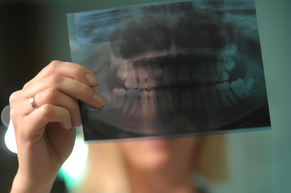
![Digital radiography technology allows dentists to take several X-rays of patients' teeth without exposing them to dangerous levels of radiation.]() By: Vojtech VlkDigital radiography technology allows dentists to take several X-rays of patients' teeth without exposing them to dangerous levels of radiation.
By: Vojtech VlkDigital radiography technology allows dentists to take several X-rays of patients' teeth without exposing them to dangerous levels of radiation. -
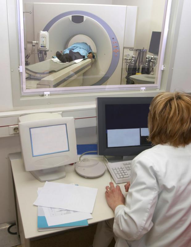
![A digital radiography technician will check that an X-ray is of high quality and depicts the body part in question.]() By: picsfiveA digital radiography technician will check that an X-ray is of high quality and depicts the body part in question.
By: picsfiveA digital radiography technician will check that an X-ray is of high quality and depicts the body part in question. -
![No evidence exists that a patient will have sustained damage from exposure to secondary radiation, such as from X-rays.]() By: labelverteNo evidence exists that a patient will have sustained damage from exposure to secondary radiation, such as from X-rays.
By: labelverteNo evidence exists that a patient will have sustained damage from exposure to secondary radiation, such as from X-rays.


Discuss this Article
Post your comments