At TheHealthBoard, we're committed to delivering accurate, trustworthy information. Our expert-authored content is rigorously fact-checked and sourced from credible authorities. Discover how we uphold the highest standards in providing you with reliable knowledge.
What is Dextrocardia?
 Tricia Christensen
Tricia Christensen
Tricia Christensen
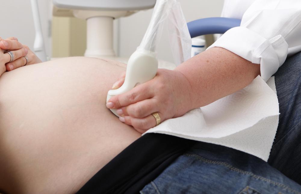
Dextrocardia is an abnormal congenital positioning of the heart in which, instead of the heart forming in the fetus on the left side, it flips over and forms on the right. There are several types of this condition, also called looping defects. It is frequently diagnosed in a routine prenatal sonogram, although not every radiologist will catch it, particularly if there are no cardiac structural abnormalities.
Mirror image dextrocardia is a very rare condition, present in about one in 130,000 people. In this looping defect, not only does the heart flip the wrong way and develop on the wrong side of the chest, but also all the other organs in the middle of the body are reversed. In essence, an X-ray of someone with this problem looks like a mirror image of the normal heart and organ placement.

This abnormality was first noted in the 1920s, when X-rays revealed this abnormal placement. Fortunately, this type of looping defect does not involve structural abnormalities of the heart or other organs. In some cases, cilia — the tiny hair-like structures — in the nose and lungs move in the opposite direction, causing a greater susceptibility to colds or illness. Aside from this susceptibility, a person with this condition does not require any special treatment or surgeries.

Dextrocardia with abnormal heart is a far more serious condition, which usually requires one or more surgeries to address structural abnormalities. It is a more common condition than the mirror image abnormality, and in most cases, the position of the other organs in the middle of the body is not reversed. The outcome and survival rates for this problem depends upon the severity of the defects, which generally include a complicated form of transposition of the arteries, called levo-transposition, or both arteries arising from the right ventricle, called double outlet right ventricle.

Large ventricular septal defects are also common in those with this condition. The right and left ventricle may be so similar, unlike in the normal heart, that it is difficult to differentiate which ventricle is which. In many cases, the ventricular septal defect is so large that these chambers are considered a single one, and one or both ventricles may be smaller than usual.

With complicated transposition and single ventricle, two surgical approaches are possible. If the ventricles are large enough, surgeons may elect to perform a Rastelli, one and a half ventricle repair. This surgery constructs a tunnel through the heart to properly reroute the blood. If possible, the ventricular septal defect may be repaired. Prior to this treatment, however, the superior vena cava is directed to the smaller pulmonary veins, and blood from the upper body never passes through the right ventricle. This initial procedure, called the Glenn shunt, reduces the heart’s workload and buys time for the child to grow before performing the Rastelli.

The Rastelli has an excellent survival rate, once past the initial surgery. It is a complicated and often lengthy procedure, with the main disadvantage of it being the length of time a child may need to be on heart lung bypass. A parent should ask a surgeon about his or her experience and success rate with this or any other surgery. Further surgical intervention may be necessary in the form of pulmonary or aortic valve replacements several years after the initial Rastelli.
Levo-transposition, small ventricles, and a high degree of pulmonary stenosis in dextrocardia with abnormal heart generally requires the three-staged Fontan operation. In this case, the surgeon performs three surgeries to palliate the defects. Improvement in the Fontan has led to improved survival rates, and many children born with this condition live well into their 30s before ultimately needing a heart transplant, the only true fix for single ventricle.
Dextrocardia may also be present in a condition called heterotaxy, which involves not only abnormal heart placement and structural abnormalities, but also the absence of a spleen or the presence of a number of small spleens. Treatment depends upon the degree and severity of these structural anomalies. Lack of a spleen, asplenia, can complicate any operations because natural resistance to infection is significantly reduced.
Tricia Christensen
Tricia has a Literature degree from Sonoma State University and has been a frequent TheHealthBoard contributor for many years. She is especially passionate about reading and writing, although her other interests include medicine, art, film, history, politics, ethics, and religion. Tricia lives in Northern California and is currently working on her first novel.
Learn more...
Tricia Christensen
Tricia has a Literature degree from Sonoma State University and has been a frequent TheHealthBoard contributor for many years. She is especially passionate about reading and writing, although her other interests include medicine, art, film, history, politics, ethics, and religion. Tricia lives in Northern California and is currently working on her first novel.
Learn more...AS FEATURED ON:
AS FEATURED ON:

-
![Dextrocardia can usually be detected in the fetus during a sonogram.]() By: Sven BährenDextrocardia can usually be detected in the fetus during a sonogram.
By: Sven BährenDextrocardia can usually be detected in the fetus during a sonogram. -
![With dextrocardia, the heart flips and forms on the right side of the fetus.]() By: Oleksandr BondarWith dextrocardia, the heart flips and forms on the right side of the fetus.
By: Oleksandr BondarWith dextrocardia, the heart flips and forms on the right side of the fetus. -
![The Rastelli procedure to fix dextrocardia is a long and complicated surgery.]() By: Tom WangThe Rastelli procedure to fix dextrocardia is a long and complicated surgery.
By: Tom WangThe Rastelli procedure to fix dextrocardia is a long and complicated surgery. -
![Dextrocardia is an abnormal congenital positioning of the heart in which, instead of the heart forming in the fetus on the left side, it flips over and forms on the right.]() By: stockshoppeDextrocardia is an abnormal congenital positioning of the heart in which, instead of the heart forming in the fetus on the left side, it flips over and forms on the right.
By: stockshoppeDextrocardia is an abnormal congenital positioning of the heart in which, instead of the heart forming in the fetus on the left side, it flips over and forms on the right. -
![Dextrocardia with abnormal heart is considered a serious condition.]() By: Patricia MarksDextrocardia with abnormal heart is considered a serious condition.
By: Patricia MarksDextrocardia with abnormal heart is considered a serious condition.


Discussion Comments
I have HLHS with dextrocardia and have been through all the fontan operations, catheters, and several pacemaker operations all starting at 2 months old and I'm about to turn 30. I've had a difficult life living with all this, but I've survived and believe anyone can survive it as long as they stay strong. Just remember life throws curve balls, but it's not hard to survive just believe in yourself and you'll be fine!
I'm 51 and went in for a chest X-Ray for back pain. Found out that I have dextrocardia. No other details. Should I be worried? Because I am.
I'm 19. I was 14 when i found out I have dextrocardia. I don't have any health issues. I play soccer and live my life like a normal person, but the only thing I'm worried about is my heartbeat. It's way slow, but normal for me, I guess.
I have dextrocardia which I found out when I was 10 years old. The location of my appendix is unknown. I am 59 years old and am fit and healthy. While in my 30s, I ran in two marathons. I am prone to chest infections, though.
I think a right hand side heart makes a person different from others. They have good abilities and are lucky.
I am 43 and also have dextrocardia. I found out when I was 10. I have not had any problems with my heart so far and I have a healthy son who just turned 18 and he was born without any problems. The only medical issue he has is a heart murmur.
I am also situs inversus totalis (dextrocardia) and I am suffering from lower abdominal pain and side pains. Can anyone tell me what I should do?
I was born with situs Inversus, third degree heart block and four holes in my heart. I also have a pacemaker. I had one open heart surgery and five surgeries to replace the pacemaker.
I'm 22 years old and other than that, I am healthy as a horse, my dextrocardia is the least worry about my heart. I have never had any problems with it and I live a happy normal life.
I believe that the way you feel about yourself and how you handle your condition, especially as a child relies wholly on your family. My family was amazing. They never treated me differently and for that I am grateful.
My wife and I just had a second degree ultrasound and the doctor told us that our baby girl's heart, stomach, and liver is mirrored. The heart is in the right place (left side chest) but turned a different way and her stomach was also mirrored, as well as her liver on the opposite side of her body going to her midline. Has anyone ever heard of this?
My wife and I are so confused and worried right now. Please let me know. --Nick
My name is Melissa and I am 26 years old. I have dextrocardia with abnormal heart, levo transposition, ventricular inversion, mitral valve regurgitation and pulmonary stenosis. I had an ASD repair when I was two and have yet to have any other surgeries or problems since. (Knock on wood). I am seeing my cardiologist tomorrow regarding becoming pregnant. I am hoping this goes well. I am active and exercise daily, eat healthy and try to manage my stress. Any advice?
@anon99240: I am also pregnant and have dextrocardia and I have seen genetic doctors and doctors who specialize in high risk pregnancies and all have said that a women who has dextrocardia can have a normal pregnancy without complications.
If you are still having concerns, going in to see your cardiologist is a way to ease your concerns also. As you get farther along in your pregnancy, they will want to monitor you and the baby closely, just to make sure the baby's organs are developing correctly.
If you do experience any chest pain or shortness of breath, then I would call your doctor to see if you need to go in. I have had a couple of chest pains and episodes of shortness of breath and they were due to my iron being low and not related to my heart. I wish you well with your pregnancy.
I am 21 years old and I was born with ASD, dextrocardia sinus inverses totalis, and pulmonary stenosis. I had heart surgery to fix the ASD when I was two months old and I am waiting to have surgery to fix the pulmonary stenosis.
The doctors told my mom I would not live past the age of ten and if I did, I would have had a dozen surgeries before then. To this day, I have only had two. I have always known my heart and organs were different from everyone else's and I have enjoyed every minute of it because I can play tricks on my friends and the doctors because it is not every day they have a patient whose heart and organs are on the opposite side.
I think of those who do have this condition to be very special and unique. I am currently pregnant with my first child and I will have a healthy little girl with her heart and organs all on the correct side.
For anyone who has this condition, it is not the end of the world, and with technology advancing all the time, there is hope for individuals who do have dextrocardia.
My son was born with situs solitus dextrocardia. His age is 11 months. Please give me your suggestions.
Well, I'm having a little trouble understanding my situation here. I'm 22 and I was born with dextrocardia, and from my understanding this is not a problem as long as it is "mirror dextrocardia", that is also what I took from this article.
Where my question is, is I do not have "mirror dextrocardia" but never had any surgeries for it. In my case, as far as I know, everything hooked up as it should have and the only thing out of place is my heart. Is this possible, or am I shooting in the dark here?
I am a 41 year old female and i found out i was dextrocardia when i was 10 and have been great. I played softball in school and still play on a co-ed league now. I had a son and the only problem he had was a heart murmur. I have been so blessed with my life. I think God every day for my life and for my health.
my sister is about 16 or 17 weeks along in her pregnancy and just found out the baby's heart has moved to the right. She is going for test next week to see the valves and all. All other organs are in place. She is lucky to work in a very good hospital.
In the high risk maternity, they are wonderful and have been since she is nervous. I have never even heard of this and stumbled on these comments. they have been very helpful. I let her know all i have read and hope it helps with how she feels. i want to say i hope god keeps blessing all of you and i know my niece will be a special little girl.
A year ago, in my X-ray lung/chest test result, there's a term "dextrocardia" written in red ink and underlined twice. The company physician wanted to see me but I was out of the office doing fieldwork. The nurse notified me that i must see another doctor and take some tests like ECG and ultrasound of the stomach area.
I was nervous upon hearing that, so i searched in the internet its meaning and was shocked upon reading posts. Since then, i can always feel my heart beat in the right side of my chest.
One week ago, an annual medical checkup was held at the office. Two doctors were amazed upon diagnosing and confirming that i am dextrocardia situs inversus totalis. Saying that I'm so special.
Well, I really thank God for designing me this way. It's not a disease; it's just being different from most of the people but still managed to cope up with the body's unusual anatomy. We are normal but very special. Thank you Lord!
i am 27 years old with dextrocardia. i came to know this last week. I have not faced any health issues in 27 years, but the chest pain started after knowing my condition. funny. Tomorrow i am going for check up. I will post my comments tomorrow again. i want to prove again dextrocardia is not a disease. Peace.
My son was born with dextrocardia. He was born premature, only weighing three pounds.
At the age of five months, he underwent his first open heart surgery, and he is surviving with one aortic valve working. At the age of five years he underwent his second and final surgery, however, as time passes by, my son has suffered three congestive heart failure episodes and liver and kidney failure.
I have been doing everything I can to help my son understand the nature of his ailment. I am proud to say with all these issues my son was able to survive his thirteenth birthday this month. He was hospitalized recently before his birthday for liver and kidney failure and dehydration.
I thank god for allowing my son to see another year and many more to come ahead. I am constantly researching on this ailment to see what treatments are available for my son and what kind of natural herbs will help him overcome this ailment.
There are so many tests that need to be done in order to work with people with these kinds of ailments, but by the grace of god I know my son is going to survive because he is strong even though he is in pain he really tries hard. Thank you for allowing me to share my news. I am from the virgin islands and never knew there were actually so many people with such an ailment. Good luck to all. I pray for an eternal life for my son and everyone with such ailments. God bless!
I was diagnosed with dextrocardia and situs inverse today! I got to the age of 30 before anything was noted, and I picked up on it, myself!
I have been referred to a cardiologist for a echo. I am amazed to have this, and I find it fascinating. Sad, I know! I have never had any problems and had a child, so for you pregnant women, all will be fine!
i don't want to say this but i am a dextrocardia child, where my heart is on the right, but my heart is not enlarged. like many other people have dextrocardia i have difficulty in breathing and i can't sleep very well.
But i thank God even though i have this, because he gave me a chance to see the world.
I am curious how many of you that are diagnosed with Dextrocardia, don't have situs inversus (mirror image), but have no other issues outside of the heart being shifted to the right.
I am in my 28th week of pregnancy. At my 20 week ultrasound my OB was having difficulty seeing my son's heart. Three weeks later, he did another ultrasound and still could not see the heart. This was all due to his positioning (head down, back facing out.) So, he sent us to a perinatologist for a more detailed ultrasound, which we saw at 25.5 weeks. That ultrasound tech was not able to see enough of the heart, also due to positioning. They did find that I had a a shortened cervix and sent me to L&D to be checked in and monitored due to possible impending preterm labor. I was transferred to a larger hospital who could accommodate my son if he was born at 25.5 weeks.
While I was in the hospital, I had three more ultrasounds, one lasting two hours and the others around an hour. None of the techs were able to get a good enough view of the heart but after the first one, the doc said that his heart appeared to be shifted to the right, close to the center of his chest and rotated 10-15 degrees further than the normal 45 degree rotation. I was discharged from the hospital and scheduled for a fetal echocardiogram the following week. At the fetal echo, the doc came in and showed us the extra rotation of the heart and said it looked like Incomplete Dextrocardia. He said that he was not able to see any other issues with his heart but was going to send us to a pediatric cardiologist to do one final echo, just to make sure they did not miss anything. The doctor told us that if there were no other concurrent medical issues or heart defects, our son would function normally and would not have any related issues, as it would just be his anatomical build. We are currently waiting for our final echo at 31 weeks.
Has anyone else had an experience like this or anything similar? My husband and I are having difficulty dealing with the not knowing and it is even more difficult not being able to find much information on the spectrum of disorders associated with this as well as the prognosis for those specific disorders. Thanks so much!
I am a 19 year old with normal dextrocardia which means all my other organs are in their normal positions. Lately i have this problem of chest pain and breathlessness. is this problem because of my habit of smoking or its due to dextrocardia? I've been paranoid the last few days. i had a chest x-ray recently which was normal. can somebody help me in knowing what's going on with me?
I'm 17 years old and i have dextrocardia with situs inversus and kartagener's syndrome. it has affected me a lot. I've not been able to go to school and i have trouble breathing. my organs are all switched and everyday life for me is hard.
Does anyone know how dextrocardia with situs inversus affects the brain?
I'm 20 years old. i was 17 when i found out that i have dextrocardia, but i have not gone to medications for my heart. i guess i am normal.
I am a 29 year woman with Dextrocadia, but all the other organ placements are normal. I have no complications other than the odd severe chest infection. I have a son and had no complications during pregnancy. I live a normal and healthy life.
My son has dextrocardia. It was discovered when he was only a few days old. The doctors told us not to worry and that it wouldn't affect him at all. He's 8 now and has never had a single complication. He plays sports, exerts himself, and is your average over-active boy.
If the doctors tell you your specific case is nothing to be concerned about, please let that relieve any fear you carry regarding dextrocardia. In most cases it's nothing that will alter one's quality of life in any way.
my granddaughter had a slight cold and was admitted to the ER and then her heart beat went up to 257. This is when the doctor discovered that she had dextrocardia at the age of three and half months old. We didn't find out about this condition until she went to the hospital. Upon her discharge from the hospital, she now appears normal and a happy baby. Nothing seems to be bothering the baby.
I have dextro and i am 31 years old with two children, eight and four years old. I have lived a normal life.
i am Enosh Wilson 29 years old. i have my heart on the right side and i am hale and hearty. i am happy i am one in a million.
I'm a healthy 28 year old female and i was diagnosed with dextrocardia when i was born. All my other organs are in the right place and I've had a very happy and normal life.
I recently found out I was pregnant and I'm concerned that my dextrocardia will make me "high risk" and what that will mean.
I was curious to hear from any women who have dextrocardia and have gone through pregnancy - is there anything I should be concerned about? Thanks!
I am a 49 years old woman with three kids all in their 20's and was only found recently to have dextrocardia, two weeks ago actually with a congested heart valve. This was only discovered by accident, (a student doctor). Life's been fine. Been, done and continue to do and go wherever I want. We're just a little out of the ordinary. good health to all.
I am 44 with mirror image dextrocardia and would like to become a donor - but would my organs be of any use? I am happy and healthy but occasionally feel a little different!
I am A 64 year old male with Dextrocardia and Situs inversus, Never had any serious problems other than respiratory infections. For all the young ones out there, it's not the end of the world. You will have a long life, just a little different from everybody else. I always tell anyone that when i was made, someone went on a break.
My son was born with dextrocardia along with other heart problems. His heart condition is called tricuspid atresia, where he only has three valves. The doctors found this out when I was pregnant with him. He had to have three open heart surgeries, with the first being at three days old. The second was at three months and his last one at four years.
I try to let him live as normally as possible with some restrictions on sports. Other than that, he is doing well. He just turned 10 in June, even though the doctors said kids with his condition don't live past the age of five. He is doing so good that they wanted to do research on his heart to see why he is doing good.
The only thing that I can tell anyone with a sick child is to pray every day. Let your kids live normal lives, because if something happens to my son tomorrow, I don't want to say that he wasn't happy while he was here.
My son was born with Dextrocardia. The doctors say everything is perfect and everything else is normal. when i first found out i was scared but i thought to myself my son is very special. wow. i have a child with his heart on the right side of his chest.
My 13 year daughter was just diagnosed last week. She has always been very healthy with no issues at all. Despite that, I am very nervous, though the doctors have told me her heart has no abnormalities at all. I can't seem to stop thinking about her condition. I am hoping that, given time, I will accept the fact that everything will be OK.
I wouldn't worry about what people say either. So what your different. God didn't mean for us to all be the same. My son is different too and as far as I'm concerned he's perfect and so are you.
I'm 14 with this and I'm a little scared because its not normal and apparently causes a lot of problems. My dad had a very small heart, but he didn't get sick. Everyone calls me the abnormal kid. But besides being a little scared I think it's really cool to be different. I don't worry what people say. They're just haters.
My sister had dextrocardia with situs inversus, and she recently died of complications with a lung infection. She was sick for many months. She was 39 years old and she lived in Honduras.
We always knew she had a "heart problem" but were not sure what it was. I think they found it when she was about 18.
Her doctor told us that she had "weak lungs" due to the mirror image. She had a very normal life until she got sick with TB, and was unable to get better. They tried a lot of different meds, but she was unable to get better.
But the doctor also told us that if she would have never gotten TB, she would not have any problems. He also said that people with mirror image have long healthy lives.
And like the WG writer stated "is mostly a curiosity more than anything else".
I hope to hear about your baby. So please write a comment after it's born. And to the 53 year old male with dextrocardia i am so happy to hear from you! It makes me believe that my son will be just fine. He's very healthy so i have no worries. But im glad that there are more people out there that are like my son. God Bless everyone that has made comments about Dextrocardia!
Thanks for your replies. It makes me feel positive that my baby will get through this and live a normal life and doesn't make me feel so alone as obviously don't know anyone else that's going through this as it's so rare! i will keep you informed and post a comment when my baby is born, hopefully it will all be good news. take care.
I am a 53 year old male with dextrocardia from birth, i have had no problems with it. I was even allowed to enter the USMC and retired from there. I hope you all do well.
My son is doing great! He also has other deformities. His aorta is too small, and he was born with two holes in his heart. He has never had a heart surgery because he is healthy.
One of the holes has healed and his heart Doc is very positive that the other one will too. He was born without ears and has congestion scoliosis. And all of his ribs are fused together on the left side and that lung is too small because of it.
But he's a healthy, totally normal two year old and i am so blessed to have him! I knew things were wrong when i was pregnant but i didn't let it bother me. I dealt with it after he was born and it wasn't so bad. There's no use in getting upset while your pregnant because the baby can feel it.
Try to tell yourself that everything will be OK. And you know what? it probably will be. Hope i get to hear from you after your baby is born. :-)
Here is a story for all other parents or parents who are expecting a child with dextrocardia.
My son was born with dextrocardia and we had no idea that anything was wrong until he was a day old. Luckily, with my son dextrocardia was not the problem. Unfortunately, he also has several other defects in his heart, which is common with dextrocardia.
He was born with an atrial septal defect, ventricular septal defect, PDA, MVP (one of his valves doesn't work right), and hole in his aorta. When he was born he was given two weeks to live.
Now for all those who are about to cry, here is the miracle. He never had a single heart surgery in his life, mainly because where we live no surgeon knew what to do with a heart affected by dextrocardia. Thankfully we would not give up and I am overjoyed to share with you all that my son will be four years old this June.
Yes, he cannot do everything other kids his age can and he tires easily, but he is in generally good health and most important of all he is alive. I hope this helps all of you that are having a hard time with sick babies.
Believe me, I understand your worries and your grief. Good luck to all of you and I hope all of you can experience the miracle my family has been blessed with.
Thanks for the reply snickers75. How is your son? Is he doing OK? Have you been told he will just lead a normal healthy life? We are obviously worried about our unborn baby and just want it to be OK!
My son has Dextrocardia. His heart was pushed over to the right side and all his other organs are where they are supposed to be. Situs Inversus is when all major organs are the opposite side and are mirror imaged. Hope this helps.
Dear anon69592: my baby girl has the same thing. It's called situs inversus. A person can live with that. my baby has to have three open heart surgeries. She had one when she was nine days old and we go back for the second one in a week, but it's because she has hypoplastic left heart. mirror image heart. And i think I said that right. but so far she is doing great. just make sure you keep the Lord first and pray. Pray a lot.
I'm 21 weeks pregnant and was told at 18 weeks that our baby has dextrocardia, that its heart is on the right hand side and is a mirror image. All other organs are in the correct place. Is this still dextrocardia or is it situs inversus. I am confused? If anyone can help, please reply.
My Husband was born with a rare form of dextrocardia, wherein his heart is on the right but all his other organs are in their correct places. He also has no sternum, and a rare form of Poland's syndrome (no pectoral muscle).
He is 35 and it has never stopped him doing anything, including playing rugby, and running his own tree surgery business. I have heard of dextrocardia but I have never heard of anyone having the same as my husband, and would love to hear of anyone else with this form of the condition.
my daughter was born with dextrocardia with abnormal heart. her doctor said she would have to have a pacemaker by the time she was five. she just turned 12 in jan and is doing great. she has had no major surgeries and only one heart cath to close one of her two holes that are in her heart. so moms it's OK. if the doc says they doing good trust in them and always keep your prayers strong. God bless each and every one of you who have this condition!
I'm 26 years old and have dextrocardia. This was discovered when I was only eight years old. I'm living a normal life with no complications whatever. I am proud to be unique.
The most funniest thing happened recently, I had a health checkup: the guy who took X-ray for me got confused. Similarly During the ECG the graph was totally reverse, the person got mad and then teared the whole graph. After swapping the ECG plugs the graph came perfect. Don't worry its not a problem. "God bless you all."
I am so happy to hear that most of the people here have had no problems with dextrocardia! My son is doing great! He hasn't had to have any kind of surgery because of his heart. His heart doctor says that he has a very healthy heart and now is only going to be seeing him once a year.
Thanks to everyone for their comments and may God be with you.
I am really happy to read these comments. One of my friends has dextrocardia. But he has not any had problems because of that. He is leading a good and healthy life. I hope he won't have any problems because of that.
My lover has dextrocardia. I was shocked when i hears it. but he has never faced any problems because of that. He is leading a beautiful life, thank god.
l have mirror image dextrocardia. i'm 38 years old and have no complications whatsoever.
my son is born with dextrocardia with situs inversus. we came to know this when he was 9 months old during his routine checkup. he is leaving normal life without any restrictions. will there be any problem in future?
One of my sister's daughter has dextrocardia. She is now eight years old and has a hole in heart as well as lacks one kidney. We have consulted many hospitals. They said no surgery will help for this. We are concerned about this and need assistance.
hi i am 25 year male suffering from dextrocardia with vsd defect. i'm a consultant designer and a civil engineer.
i have faced many critical problems during the last 10 years but take some precautions with homeopathic medicine and i live my normal life.
I am a 31 year old woman who was born with dextrocardia as well as a hole in my heart which has mended itself. Please do not fear this as i have lived a normal life with no restrictions. At eight years old my doctor told me to leave the office and to live normally. I went back at 23 years old to have him explain everything to me and again he told me i am to live with no restrictions. Please know that this is a special part of me, it's rare and for that i know i am unique. Those who are pregnant with a child with dextrocardia please know that this does not mean you have a disabled child by any means. God bless you and your family.
Dear Iluvg8tr for your comments.
I am 21 weeks pregnant and was told at 18 weeks pregnant that my baby girl has dextrocardia with situs inversus. My husband and I had not heard of this until then and naturally very concerned about the implications on our baby's health. We have seen a children's heart specialist since, who says that her heart is functionally normal despite the dextrocardia, and have been told by doctors that she can lead a normal healthy life but we cannot help but still we worry. Your comments made me feel so much better, so thank you.
I am 23 weeks prego and am having a little girl with dextrocardia. I live in a small town in texas where my doctor has never seen this before, so they have shipped me all over. I will soon see a doctor in Fort Worth and hope for the best. I see all the comments and it makes things a little easier for me. thank you and god bless all the other families for their input.
Thank you everyone for the kind words and reassurence. I think my son will be just fine. He's very healthy and hasn't had to have surgery on his heart.
I was born with dextrocardia and situs inversus, thanks to god i did not need any surgery. I am 42 years old, married and have two beautiful children without the same condition (early screening during pregnancy was performed).
I have lived my life without any restrictions or limitations, just recently got some bad cholesterol but it has been controlled.
I remember jokes with the medical interns, the doctor put the electrical contacts during routine checks (electrocardiogram) and everything was inverse in the printings, the interns scratched their heads when asked what is wrong, I laughed.
I write this to let you know that life goes on, god bless you all.
I'm a seventeen year old with mirror image dextrocardia. I play volleyball and live a very normal life :) Though it may be scary at first; put your faith in the Lord. --Ashley, KY
Dear Jill61945, I am so happy to hear about how well your grandson is doing!!! Sounds like he is getting a better start in life. Keep us posted. Id like to hear more. My son hasn't had to have any surgeries but I do know about the heart issue because my sons heart is on the wrong side and has a few more problems. But I would love to hear how your grandson is doing every once in awhile.
Dear anon31347, I have given birth to four healthy grown up kids.
Hi. Im 51 years old and a dextrocardia. This was discovered when I was only 4 years old. Im living a normal life with no complications whatever.
Im having a baby girl in Sept.. im 25 weeks and last week the doctors told us that her heart and stomach was on the right side. they said she has Dextrocardia but she also has 2 holes in her heart or sumthing called Atrioventricular septal defect and she has Heterotaxy syndrome and abnormal visceral situs.. They said she might have 2 have surgery her 1st couple of years.. has anybody have sum advice or ever heard of sumthing like this....?
My daughter was Dextrocardia with Situs Inversus, meaning she is a mirror image of all of her organs including her heart. We didn't find out until she was 18 months old during a routine Dr's visit. She had a cold and when we took her to the doctor they did a chest xray and this is when they discovered it. Sounds odd that they wouldn't know when I was pregnant but there was no reason to suspect anything.
She spent 1 week in the hospital after birth for jaundice and at 14 months old spent 2 days in the hospital for the roto-virus. During those hospital visits they didn't discover it then either. Shortly after finding out she went to Children's Hospital in Atlanta for a bronchial study to see if she had Kartagener's syndrome. Luckily she doesn't! She is a healthy thriving 3 year old little girl that really the only complication we have with this is some breathing issues like Asthma. She takes singular and Qvar just like any other child takes for Asthma. She is having her tonsils and adenoids taken out in July because she snores so loudly. With her lungs being reversed she already has a little bit of a challenge so we are hoping this surgery will help her to breathe better and sleep better.
My husband and I realize that we are one of the lucky ones since all of her organs mirrored. There are times it does get frustrating having to explain her condition to everyone from daycare, to sitters, to just the average person who is curious. She sees her pediatric cardiologist once per year just to check her heart but everything appears perfect although backward. She is truly a miracle and I wouldn't trade her for anything in the world.
If you have a child born with this condition just know that there may be a few more doctors that are curious and want to "see" for themselves through x-rays. There may be a few more boxes to fill out on a health screen questionnaire, and a few more co-pays and deductibles to pay because of having to see a specialist for even the slightest little thing but embrace your child's difference and know that having a child as unique as this is truly a blessing. If anyone ever needs to chat about dextrocardia I'll be happy to be your friend.
I wrote about my great-grandson when he was 6 months old having dextrocardia and a mirror image heart. He is now 9 months old and just had his second open heart surgery to re-route an artery and to remove the shunt that was put in when he was 2 weeks old. He is doing really well and if you didn't know it, he looks and acts like any other baby. The only noticeable trait is his little fingernails are a little blue, but other than that he is fine. He plays, he crawls a little, and eats good. He has two teeth and three more are coming in. He will have to have one more surgery to correct the arteries when he is about 4 years of age.
I just want to let anyone out there know that if your child has this condition, or you know somebody who has dextrocardia that it can be treated and you can live a relatively normal life. They did tell us that Cole may not live to be an "old" person without a heart transplant, but with technology these days, who knows what new procedures will be available later on.
What problems may I face in my marriage having dextrocardia?
Dear everybody,
My two sons have a case called kartagener syndrome with dextrocardia.Tthey have health problems.
I want to know is there a permanent cure .
Dear Jill61945
Your grandsons oxygen level doesn't sound right to me, My sons heart doctor says that my sons oxygen level needs to be in the 90s. I suggest that you get a second opinion. No baby gets used to having low oxygen. I hope this helps you. May god be with you and your grandson.
My great-grandson who is 6 months old has dextrocardia and two holes in his heart. He has had one surgery and is scheduled for another next month. My biggest worry right now is that his oxygen level is dropping. Right now it is in the low 70s. Otherwise, he is a happy baby, he is growing, teething, and seems normal in every other way. Sometimes his hands and feet look bluish. The cardiologist says he is adjusting to this lower oxygen level but I am wondering what effect that will have on the rest of his body before he has all the surgeries he needs to correct the problems.
Hi, My sister had dextrocardia, but unfortunately she died when she was 6 due to a very sudden and severe chest infection. It was a very long time ago, when information and knowledge were scarce about this condition. My parents were not really aware of the conditions and complications they should look out for. Fortunately, these days, there is plenty of information available. I hope that everyone who is affected by this condition gets all the facts that they need.
Thank you wgwriter for your kind words. It helps so much to know that there are more people going through what im going through. Hope all is well on your end too.
Snickers, That's terrific that he's doing so well, and as mentioned some holes do repair themselves and never need surgery. Sticking with cardiologist advice is just good sense on this. It's always a good idea to have an exact diagnosis (if its hard to remember have your cardiologist write it down for you), just in case a child needs emergency care. Preemies are much more prone to heart defects that do close, particularly holes in the atrial septum and a continued opening of the patent ductus. I'm sure the book, and continued doctor's advice will really help you out on this, but each kid's case is different. I'm so glad to hear he is healthy and doing well, though I know it can be challenging raising a baby with any problems :)
Wow! Its Crazy but i don't know where the holes are exactly in his heart. All i know is that his heart doctor said that they are fixing themselves and that she can hardly hear them any more. And that she doesn't think that he will need surgery in the future. And when it comes to his weight gain he's doing so good. He is a preemie so of course he's smaller than a normal baby his age but he just keeps on putting on the weight. Im very proud at how well he is doing and so are all of his doctors because he has more then just a heart condition. He also has congenital scoliosis and his ears never developed. For right now he's as healthy as a horse. He sees his heart Doctor in September but i will call the office and find out about the holes and let you know.
Snickers, Can you elaborate on the holes in his heart? That could suggest some other conditions. Is your son dextrocardia with mirror image placement of the organs, or another condition related is dextrocardia with abnormal heart.
Generally, unless the holes are located in the patent ductus area, they may require surgical intervention at a later point. Keep me posted on this. I assume you mean a few holes in the septum (the dividing wall between the two sides of the heart)? Sometimes this can even be fixed now by non-invasive methods, but it can cause problems in infants, especially in the areas of gaining weight properly. Many kids can go for years without needing surgery, but I'd like to know specifically, if you don't mind, what the doctors are thinking about your son. Note I am not a doctor (nor do I play one on television), but I did run a support group for families with kids with heart conditions for 5 years. I have a bit more experience in this area given my son's condition.
Thank you anon13937 for posting a comment about dextrocardia. It brings me great hope that my son will be OK. He also has a Bicuspid Aoritic Valve and two small holes in his heart but his heart doctor says that he is doing great. And too WGwriter, I will read the book The Heart of a Child. Thank you.
Hi, I'm twenty and I have Dextrocardia.
I love to tell people that my heart is on the other side of my chest! I am a perfectly healthy male. I also have a Bicuspid Aortic Valve. I really hope that doesn't cause problems later in life.
Hi Snickers75,
My son has dextrocardia too, though he had a lot of other heart conditions that required several surgeries. From what I know about mirror image dextrocardia, there are a couple of complications you may look for. First, he should have an emergency bracelet. If your son ever needed emergency surgery, you'd want to make sure a surgeon was operating in the right place and not making unnecessary incisions. Second, the cilia, fine little hairs in the nose and lungs, tend to beat in the wrong direction. This may make your son a little more susceptible to colds and flus. You might want to be sure your son gets flu shots each year.
Generally, though, when no other abnormality of the heart's structure exists, your son should do very well. It's a medical curiosity more than anything else, and most people with only the mirror image condition lead very normal lives.
You might want to obtain a copy of "The Heart of a Child" a very good book that explains lots about heart conditions in general.
my son was born with mirror image dextrocardia and everything else seems to be normal. My question is, will he have complications in the future?
Post your comments