At WiseGEEK, we're committed to delivering accurate, trustworthy information. Our expert-authored content is rigorously fact-checked and sourced from credible authorities. Discover how we uphold the highest standards in providing you with reliable knowledge.
What is an Evoked Potential?
An evoked potential (EP) is an electrical signal that is recorded or generated from the central nervous system in response to the presentation of a stimulus. Evoked potential tests are conducted on patients who may experience sensory deficit and are unable recognize a stimulus. Evoked potential signals can be recorded from a variety of areas, including the cerebral cortex, spinal cord, and peripheral nerves. This is distinct from the spontaneous potentials that are detected by electroencephalography (EEG) or electromyography (EMG). Visual, auditory, and somatosensory stimuli are commonly used for evoked potential studies.
The individual performing an EP test locates specific areas on the patient's head to place electrodes. For a visual evoked potential, the patient focuses on a screen that displays a checkerboard pattern, with one eye covered with a patch. Auditory evoked potentials involve headphones that are used to deliver a series of clicks to one ear, while a static sound is played into the other ear. For somatosensory EPs, electrical shocks are delivered to the arm or leg, causing some tingling.

Somatosensory evoked potentials (SEPs) deal with waves that are reflected by the activation of neural structures along the somatosensory pathways. Clinical studies generally use electrical stimulation of peripheral nerves to elicit stronger responses. The areas usually tested in clinical studies for somatosensory evoked potentials include the posterior tibial nerve at the ankle, the median nerve at the wrist, and the common peroneal nerve at the knee.

SEPs are used in diagnosing neurologic disease in patients. They are also used to monitor surgeries involving risk to the somatosensory pathways. Although abnormal results of SEPs can result from some form of dysfunction at the peripheral nerve level, patients cannot force abnormal results. Abnormal SEP results suggest a dysfunction within the somatosensory pathways.

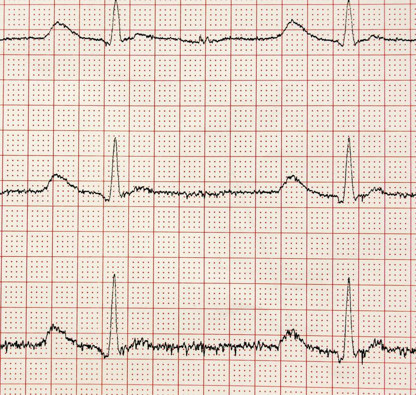
Evoked potential amplitudes tend to be much lower than EEGs or EMGs. Amplitudes from evoked potentials tend to be less than a few microvolts, in comparison to thousands of microvolts for EEG and close to a volt for ECG. Signal averaging is therefore required to resolve these low-amplitude potentials against EEG, ECG, EMG, and surrounding sounds. Since the background noise is random, it can be averaged out with the mathematical mean of repeated responses.

Clinical settings involving somatosensory evoked potentials have been greatly impacted by the development of refined neuroradiologic imaging. Although fewer diagnostic SEP studies have performed recently, SEPs are still valuable diagnostic tests in many clinical situations. SEPs are widely used as research tools for sensory physiology. In addition, somatosensory evoked potentials are becoming more important in the operating room.
AS FEATURED ON:
AS FEATURED ON:

-
![A type of neurofeedback equipment, an electroencephalogram -- or EEG -- machine measures electrical impulse activity in the brain and can help give a diagnosis for conditions such as epilepsy.]() By: satoriA type of neurofeedback equipment, an electroencephalogram -- or EEG -- machine measures electrical impulse activity in the brain and can help give a diagnosis for conditions such as epilepsy.
By: satoriA type of neurofeedback equipment, an electroencephalogram -- or EEG -- machine measures electrical impulse activity in the brain and can help give a diagnosis for conditions such as epilepsy. -
![Clinical studies for SEPs test the tibial nerve at the ankle, the median nerve at the wrist, and the common peroneal nerve at the knee.]() By: 7activestudioClinical studies for SEPs test the tibial nerve at the ankle, the median nerve at the wrist, and the common peroneal nerve at the knee.
By: 7activestudioClinical studies for SEPs test the tibial nerve at the ankle, the median nerve at the wrist, and the common peroneal nerve at the knee. -
![Evoked potential amplitudes tend to be much lower than EEGs.]() By: a40757seEvoked potential amplitudes tend to be much lower than EEGs.
By: a40757seEvoked potential amplitudes tend to be much lower than EEGs. -
![A doctor may order an EEG for patients suffering from headaches.]() By: JPC-PRODA doctor may order an EEG for patients suffering from headaches.
By: JPC-PRODA doctor may order an EEG for patients suffering from headaches.


Discuss this Article
Post your comments