At WiseGEEK, we're committed to delivering accurate, trustworthy information. Our expert-authored content is rigorously fact-checked and sourced from credible authorities. Discover how we uphold the highest standards in providing you with reliable knowledge.
What Is an Aortography?
Aortography is a diagnostic tool that uses contrast dye to evaluate arterial blood flow. Also known as an angiogram, aortography is commonly performed to assess arterial function through the aorta, which runs the length of one’s trunk from the heart down to the abdomen. Designed to produce a two-dimensional image of a targeted section of the aortic artery, there are risks associated with this form of projectional radiography, including infection.
Individuals who undergo aortography are given a local anesthetic where the catheter will be positioned. It is not uncommon to suddenly feel warm when the dye is introduced into the bloodstream. Generally taking less than an hour, individuals may spend several hours in recovery to ensure there are no complications. Within 24 hours, individuals are usually able to participate in everyday activities without restriction.

The way the test works is that a small, hollowed catheter is introduced into the groin or arm by a thin needle. X-ray is used to guide the needle to its target location within the aortic artery. Once in position, contrast dye is introduced into the bloodstream through the catheter and, as the dye dissipates throughout the artery, its progress is monitored. After a picture of the targeted aortic region is obtained the catheter is taken out and the injection site is bandaged.

Abnormal aortographic results can be indicative of a variety of medial situations and conditions. Arterial injury, narrowing or valve impairment may be detected with aortography. Congenital heart conditions, aortic aneurysm and peripheral artery disease (PAD) may also be diagnosed or confirmed as a contributing factor to impaired arterial function. Depending on results, additional testing may be ordered.

Several types of projectional radiography are performed, depending on the suspected area of impaired aortic blood flow. Blockages within the lower aortic artery, such as the groin area, can necessitate translumbar aortography, which offers a picture of the lower abdominal region. If there is a question about the health of the upper aortic artery, supravalvular aortography is conducted. A suspected blockage or other form of impaired aortic blood flow in the abdomen usually requires abdominal aortography.

Prior to testing, individuals are given specific instructions to help ensure untainted results. Medications that may interfere with blood clotting are usually temporarily discontinued. The patient is instructed to not consume any food or beverages at least six hours prior to testing.
There are risks associated with aortography. Anytime you introduce a foreign substance into the body, such as contrast dye, there is a chance for allergic reaction. The site where the catheter is positioned may bruise, bleed or become infected. It is possible for catheter placement to also impair or permanently damage the blood vessels and nerves in the immediate area.
AS FEATURED ON:
AS FEATURED ON:

-
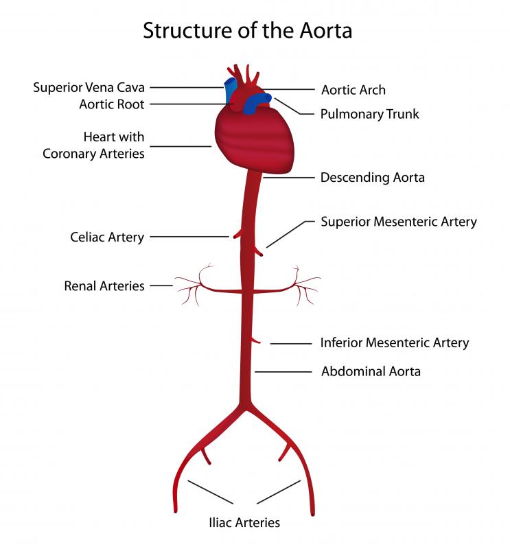
![A diagram of the aorta.]() By: bilderzwergA diagram of the aorta.
By: bilderzwergA diagram of the aorta. -
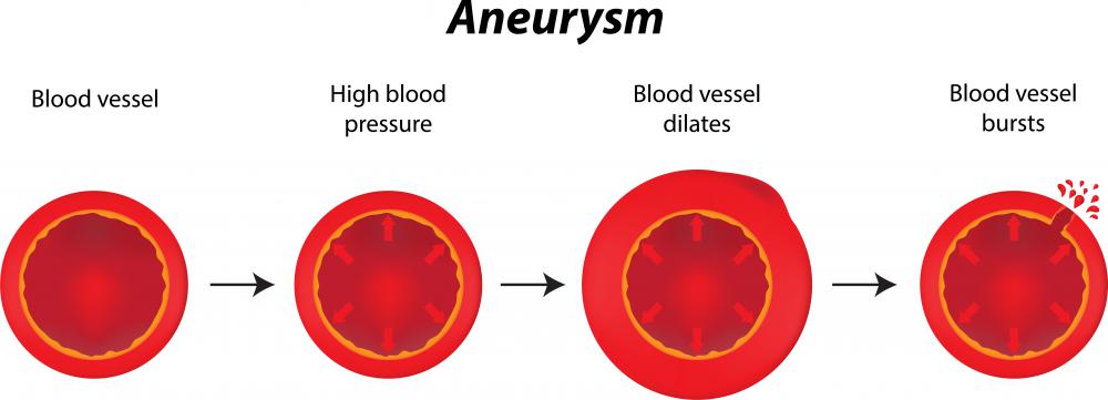
![An aortography might be used to detect an aneurysm.]() By: joshyaAn aortography might be used to detect an aneurysm.
By: joshyaAn aortography might be used to detect an aneurysm. -
![The injection site is usually bandaged following an aortogrpahy.]() By: amawasriThe injection site is usually bandaged following an aortogrpahy.
By: amawasriThe injection site is usually bandaged following an aortogrpahy. -
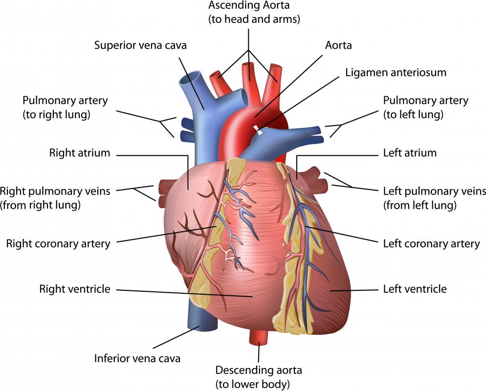
![Aortography provides information about the shape, structure, size and position of the aorta.]() By: peterjunaidyAortography provides information about the shape, structure, size and position of the aorta.
By: peterjunaidyAortography provides information about the shape, structure, size and position of the aorta.

Discuss this Article
Post your comments