At WiseGEEK, we're committed to delivering accurate, trustworthy information. Our expert-authored content is rigorously fact-checked and sourced from credible authorities. Discover how we uphold the highest standards in providing you with reliable knowledge.
What is a Radiograph?
A radiograph is an image taken with X-ray technology that allows the inside of an object to be seen. X-rays, also called X radiation or Roentgen radiation, are a type of electromagnetic radiation with a very short wavelength. The radiation with the shortest wavelengths, hard X-rays, are powerful enough to penetrate objects, making them a useful tool for security screening, medical diagnostics, and looking at the inside of crystals. The radiograph is a two dimensional picture of objects in shades of white on a black background.
A radiograph is produced by beaming hard X-rays through the subject, whose insides absorb different amounts of radiation depending on the densities of its components. In an X-ray image of the body, bones, which are dense, absorb more radiation than soft tissues, which are less dense. This absorption of radiation is called attenuation. Where more radiation is attenuated, less radiation is able to pass through to the other side of the object. Where there is less attenuation, more X-rays are able to fully pass through the subject.

The radiation that makes it through to the other side of the subject is caught by an X-ray sensitive plate. Where it hits on the plate, the radiation excites electrons, or subatomic negatively charged particles. In a photographic plate, the old means of viewing a radiograph, these exposed areas would darken, the less exposed areas would show up gray, and the unexposed areas would remain white. This is why a radiograph of the body shows the bones as white, the soft tissues as gray, and the background as black.

Today, the photographic plate has largely been replaced by computed radiography, which uses photostimulable phosphor plates (PSP plates). In this process, the radiation penetrates the subject, hits the plate, and excites the electrons in the areas where the subject is less dense. This piece of the procedure is similar to using a photographic plate, except less radiation can be used. Less radiation is preferred, as high amounts of radiation may mutate cells in a harmful way. After the PSP plate has been exposed, the electrons are beamed with a laser and the signal is then run through a computer and translated into a digital image.

In a medical setting, the radiograph is typically used to examine bones, but a softer X-ray, or one with a longer wavelength, can be used to look at soft tissues. Radiography also includes fluoroscopy, an imaging technique that achieves a lower resolution moving image of the body. This is used to examine moving tissues, such as the flow of blood, or to guide surgical procedures. X-ray technology also has many industrial uses, such as scanning luggage at the airport, viewing the inside of cargo crates, and inspecting the inside of products to ensure safety and quality.
AS FEATURED ON:
AS FEATURED ON:

-
![A radiograph is an image taken with the use of x-rays.]() By: creo77A radiograph is an image taken with the use of x-rays.
By: creo77A radiograph is an image taken with the use of x-rays. -
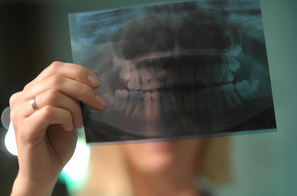
![Dentists use radiographs to check for cavities, fractures and issues below the gum line.]() By: Robert KneschkeDentists use radiographs to check for cavities, fractures and issues below the gum line.
By: Robert KneschkeDentists use radiographs to check for cavities, fractures and issues below the gum line. -
![Radiation that makes it through to the other side of the subject is caught by an X-ray sensitive plate.]() By: inkaoneRadiation that makes it through to the other side of the subject is caught by an X-ray sensitive plate.
By: inkaoneRadiation that makes it through to the other side of the subject is caught by an X-ray sensitive plate. -
![Digital radiography technology allows dentists to take several X-rays of patients' teeth without exposing them to dangerous levels of radiation.]() By: Vojtech VlkDigital radiography technology allows dentists to take several X-rays of patients' teeth without exposing them to dangerous levels of radiation.
By: Vojtech VlkDigital radiography technology allows dentists to take several X-rays of patients' teeth without exposing them to dangerous levels of radiation.


Discuss this Article
Post your comments