At AllTheScience, we're committed to delivering accurate, trustworthy information. Our expert-authored content is rigorously fact-checked and sourced from credible authorities. Discover how we uphold the highest standards in providing you with reliable knowledge.
What is a Metaphase?
Metaphase is one of the stages of mitosis and meiosis, which are the two types of cell division. During mitosis, cells are produced that are genetically identical to the parent, or clones. It is used for asexual reproduction, growth of multi-cellular organisms and for repairing and replacing damaged tissues. Meiosis is the cell division that is used to produce cells for sexual reproduction. Mitosis happens in all cells, while meiosis only occurs in the sexual organs of an organism, e.g. the testes and ovaries of mammals or the ovaries and anthers of flowering plants.
Both mitosis and meiosis are continuous processes, but they are each described as a series of stages. During mitosis, there are four stages – prophase, metaphase, anaphase and telophase, which occur in that order. Meiosis has two divisions, meiosis I and meiosis II, each made up of the same four stages as mitosis. There is one further stage for both processes called interphase. Interphase occurs before the dividing stages and it is when the cells grow and prepare to divide by replicating their DNA.

All cells have a cell cycle that starts when they have been produced through cell division and ends when they divide to produce identical cells. Mitosis is the period of cell division and the rest of the cell cycle is interphase. Interphase is commonly referred to as the resting phase, but is a time of much cellular activity. During this phase, the cell grows and produces organelles and proteins. The DNA in the nucleus is replicated in preparation for mitosis and it continues to grow and produce duplicate organelles.

During prophase, the chromosomes in the nucleus get shorter and thicker, condense and become visible. Each chromosome appears to have two chromatids that are joined together by a centromere. Centrioles form and move to opposite ends of the cells where microtubules develop to form a star-shaped structure called an aster. Some of the microtubules, or spindle fibers, cross the cell from end to end to form the spindle. Lastly, the nucleolus and nuclear membrane break down so the chromosomes are free floating in the cytoplasm.

The next stage of division after prophase is metaphase. During this stage, the chromosomes line up along the middle of the cell. Each of the chromosomes is attached to a spindle fiber at their centromere. The chromatids are then pulled slightly apart due to the contraction of the microtubules. Anaphase and then telophase follow on from metaphase.
During anaphase, the spindle fibers are completely contracted so the separate chromatids of each chromosome are pulled to either side of the cell. Once the chromatids reach the poles of the cell, a new nuclear membrane forms around them, indicating the start of telophase. The spindle fibers break down, the chromosomes uncoil and elongate, the nucleolus reforms and, finally, the cell divides into two ending the mitotic division.
Meiosis is similar to mitosis, but two divisions take place. It involves the division of the chromosomes followed by two divisions of the nucleus and cell. Meiosis I differs from mitosis during prophase, but meiosis II is a typical mitotic division, as described above. The end result of meiosis is four new cells that have half the genetic information of the parent cell.

The key difference in meiosis I occurs during prophase I when the chromosome pairs come together to form a bivalent instead of each chromosome forming a chromatid. During metaphase I, the bivalents randomly line up along the middle of the cell to be separated. This random orientation leads to increased genetic variety. Each chromosome of the pair has genes that determine the same characteristics, but they are not always the same gene. The random distribution and following independent assortment of the chromosomes creates new genetic combinations in the cells.
The chromosomes are pulled to opposite ends of the cell during anaphase I, and a nuclear membrane forms around them in telophase I. The resulting two cells now have half as much genetic material as the parent cell. Meiosis II follows the same process as mitosis, where the chromosomes form a pair of chromatids joined by a centromere. They line up along the center of the cell and are pulled by their centromeres to the opposite ends of the cell. Once they reach the poles, cell division completes resulting in four new cells, each with half the genetic material of the original cell.
AS FEATURED ON:
AS FEATURED ON:

-
![A cell dividing into two daughter cells.]() By: Luk CoxA cell dividing into two daughter cells.
By: Luk CoxA cell dividing into two daughter cells. -
![A cell.]() By: Amelie OlivierA cell.
By: Amelie OlivierA cell. -
![The stages of mitosis.]() By: VanessaThe stages of mitosis.
By: VanessaThe stages of mitosis. -
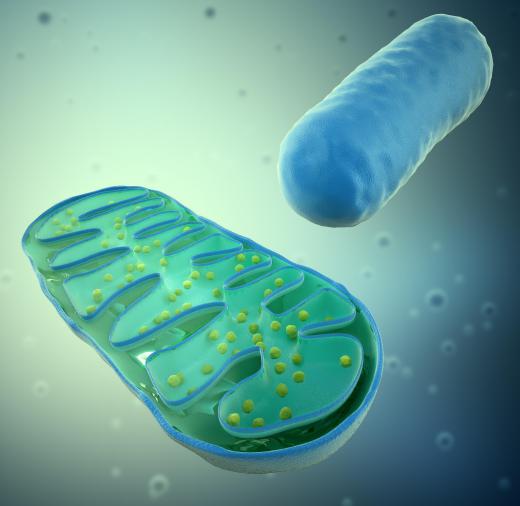
![Duplicate mitochondria are produced during metaphase.]() By: MopicDuplicate mitochondria are produced during metaphase.
By: MopicDuplicate mitochondria are produced during metaphase. -
![During prophase, the chromosomes in the nucleus get shorter and thicker, condense and become visible.]() By: Giovanni CancemiDuring prophase, the chromosomes in the nucleus get shorter and thicker, condense and become visible.
By: Giovanni CancemiDuring prophase, the chromosomes in the nucleus get shorter and thicker, condense and become visible.


Discuss this Article
Post your comments