At WiseGEEK, we're committed to delivering accurate, trustworthy information. Our expert-authored content is rigorously fact-checked and sourced from credible authorities. Discover how we uphold the highest standards in providing you with reliable knowledge.
What is a Fundoscopy?
 Malcolm Tatum
Malcolm Tatum
Malcolm Tatum
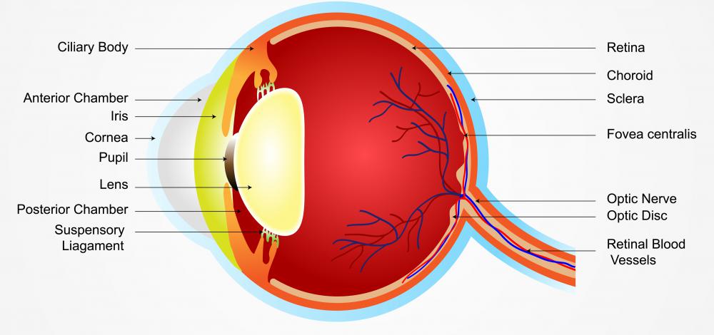
Also known as ophthalmoscopy or funduscopy, fundoscopy is a procedure in which the back portion of the eye is examined. This part of the eye, known as the fundus, includes the blood vessels that feed the eye, the retina, the optic disk, and the choroid. There are actually three different forms of this type of procedure, all of them conducted using an instrument known as an ophthalmoscope or a fundoscope.
The first version of the procedure is known as direct fundoscopy. In this process, the patient is placed into a darkened room, usually in a sitting position. A small beam of light is directed on the pupil of the eye, using the ophthalmoscope. The light is sufficient to allow the ophthalmologist to examine the back portion of the eye as the patient is asked to look in various directions.

Indirect fundoscopy involves placing the patient into a semi-recline position, and applying drops that help the eye to dilate. The eyelids are held open while a bright light is directed at the eye. With this procedure, the ophthalmologist wears a light on his or her head, often using a device that resembles a miner’s hat. This leaves the healthcare professional’s hands free to use a small tool to press lightly against the eye during the examination as the patient is asked to look in different directions.

Slit-lamp fundoscopy also involves placing the patient in a sitting position, and administering drops into the eyes. The slit-lamp includes a chin rest that makes it easier for the patient to maintain a still position during the course of the examination. With this approach, a narrow band of light is directed into the eyeball, allowing the ophthalmologist to clearly identify any conditions that indicate the presence of some type of disease or degradation of the eye. The intensity of the light used with this procedure sometimes causes patients to be light-sensitive for several hours, requiring patients to use sunglasses or other forms of eyewear to protect the eyes until the sensitivity fades.

People with diabetes often have a fundoscopy of some type performed at least once a year. Depending on the severity of their condition, this type of examination should be conducted every six months. This makes it possible to detect any changes in vision that may be occurring due to the progress of the disease, allowing medical professionals to explore options to minimize the damage to the eyes.
Malcolm Tatum
After many years in the teleconferencing industry, Michael decided to embrace his passion for trivia, research, and writing by becoming a full-time freelance writer. Since then, he has contributed articles to a variety of print and online publications, including WiseGEEK, and his work has also appeared in poetry collections, devotional anthologies, and several newspapers. Malcolm’s other interests include collecting vinyl records, minor league baseball, and cycling.
Learn more...
Malcolm Tatum
After many years in the teleconferencing industry, Michael decided to embrace his passion for trivia, research, and writing by becoming a full-time freelance writer. Since then, he has contributed articles to a variety of print and online publications, including WiseGEEK, and his work has also appeared in poetry collections, devotional anthologies, and several newspapers. Malcolm’s other interests include collecting vinyl records, minor league baseball, and cycling.
Learn more...AS FEATURED ON:
AS FEATURED ON:

-
![An optometrist uses a slit lamp machine to examine a patient's eyes.]() By: daniel rajszczakAn optometrist uses a slit lamp machine to examine a patient's eyes.
By: daniel rajszczakAn optometrist uses a slit lamp machine to examine a patient's eyes. -
![People who've just undergone a fundoscopy may need to wear sunglasses to protect their eyes from light.]() By: MiravisionPeople who've just undergone a fundoscopy may need to wear sunglasses to protect their eyes from light.
By: MiravisionPeople who've just undergone a fundoscopy may need to wear sunglasses to protect their eyes from light. -
![Normally, a person's pupils contract when light is bright, but eye doctors will usually use drops to dilate the pupils temporarily to see the back of the eye.]() By: mtthsNormally, a person's pupils contract when light is bright, but eye doctors will usually use drops to dilate the pupils temporarily to see the back of the eye.
By: mtthsNormally, a person's pupils contract when light is bright, but eye doctors will usually use drops to dilate the pupils temporarily to see the back of the eye. -
![The fundus includes the blood vessels that feed the eye, the retina, the optic disk, and the choroid.]() By: stockshoppeThe fundus includes the blood vessels that feed the eye, the retina, the optic disk, and the choroid.
By: stockshoppeThe fundus includes the blood vessels that feed the eye, the retina, the optic disk, and the choroid. -
![It's important to have regularly scheduled eye exams in order to minimize any damage to the eyes.]() By: FotolEdharIt's important to have regularly scheduled eye exams in order to minimize any damage to the eyes.
By: FotolEdharIt's important to have regularly scheduled eye exams in order to minimize any damage to the eyes. -
![A slit-lamp or ophthalmoscope is used to shine light on the back portion of the patient's eye during a fundoscopy.]() By: Monkey BusinessA slit-lamp or ophthalmoscope is used to shine light on the back portion of the patient's eye during a fundoscopy.
By: Monkey BusinessA slit-lamp or ophthalmoscope is used to shine light on the back portion of the patient's eye during a fundoscopy.


Discussion Comments
The "miner's hat" is called a Binocular Indirect Ophthalmoscope (or BIO for short). A lens is held in front of the eye during this procedure; nothing is pressed directly against the eye.
Any given optometrist should be doing both slit lamp fundoscopy and binocular indirect ophthalmoscopy each time you visit them, possibly excluding follow-up visits.
Yes, it is a pain to be dilated and not be able to read for three hours, but on the other hand, these procedures can help identify certain conditions which can eventually cause permanent blindness. Most often, this blindness is preventable. Before going to see the eye doctor, make arrangements -- pick up some cheap +1.50 reading glasses and possibly some sunglasses if you plan to be outside after dilation -- and get your fundus checked regularly.
Post your comments