At TheHealthBoard, we're committed to delivering accurate, trustworthy information. Our expert-authored content is rigorously fact-checked and sourced from credible authorities. Discover how we uphold the highest standards in providing you with reliable knowledge.
What Are White Matter Foci?
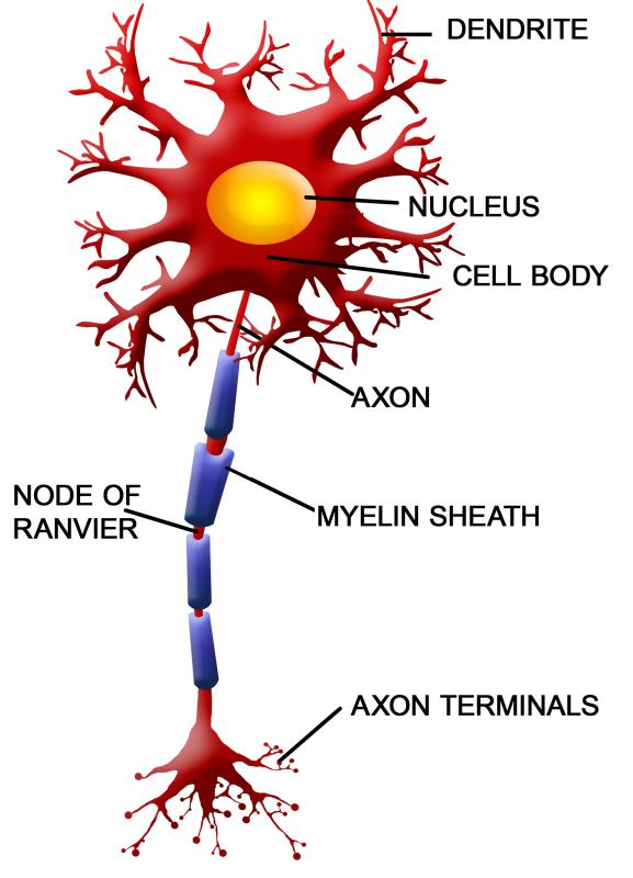
White matter foci commonly appear on a magnetic resonance imaging (MRI) as bright white spots on the part of the brain that contains nerve cells covered with lipid tissue known as myelin. The foci typically appear in areas with higher levels of fluid. They usually indicate physiological changes caused by disease processes, infections, or the normal aging process. Individuals with white matter foci may or may not exhibit physical symptoms.
People often equate these bright spots with the potential diagnosis of multiple sclerosis (MS) or brain tumors, but this is not necessarily the case. Medical professionals evaluate the spots based on a patient’s physical symptoms, the location and size of the lesions, and information gained from other tests. Proper diagnosis may require the opinion of radiologists, neurologists, or other specialists, combined with a series of bodily fluid tests and physical exams.

Multiple sclerosis involves the deterioration of the myelin sheath, which an MRI scan depicts as white matter foci in various parts of the brain. Patients with MS usually complain of visual disturbances along with numbness or weakness in the extremities. Healthcare professionals make a definitive diagnosis based on the MRI, spinal fluid tap, and extensive visual testing. Health care providers usually order MRIs over a period of time, checking for an increased number of foci and the location of plaque development.

Patients with bacterial, fungal, or viral encephalitis commonly exhibit these spots on MRI scans as well. The areas typically indicate abscesses or vascular inflammation. Patients generally report having had an infection or experiencing physical symptoms, which is why the imaging studies are performed. Diagnosis and treatment usually requires identification of the organism through blood and urine samples, spinal tap, and sensitivity cultures.

Brain tumors and post-stroke hemorrhage frequently appear as white matter foci, and MRIs with contrast media generally further indicate specific vascular involvement. Patients with either diagnosis generally experience physical symptoms prompting the imaging studies. Tumor treatment depends on whether the lesion is benign or malignant, its size, and its location. Treatment for lesions that appear secondary to a stroke depend on whether a vessel is blocked or ruptured and whether it is accessible.

Patients diagnosed with hypertension, diabetes, or high cholesterol often have white matter foci. These areas generally indicate restricted blood flow in the brain’s capillaries. These lesions frequently appear as individuals get older, and in most instances, these patients don't have physical symptoms. Healthcare professionals generally prescribe medication for the underlying cause and follow up as needed. Spots have also been discovered in the brains of patients with a history of migraines.
AS FEATURED ON:
AS FEATURED ON:

-
![People with hypertension often have white matter foci.]() By: kazoka303030People with hypertension often have white matter foci.
By: kazoka303030People with hypertension often have white matter foci. -
![People with diabetes often have white matter foci.]() By: charger_v8People with diabetes often have white matter foci.
By: charger_v8People with diabetes often have white matter foci. -
![White matter foci frequently appear as individuals get older.]() By: jamiehooperWhite matter foci frequently appear as individuals get older.
By: jamiehooperWhite matter foci frequently appear as individuals get older. -
![People with a history of migraines may have white matter foci.]() By: Photographee.euPeople with a history of migraines may have white matter foci.
By: Photographee.euPeople with a history of migraines may have white matter foci. -
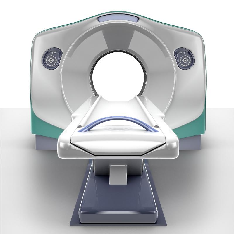
![Magnetic resonance imaging scans reveal white matter foci.]() By: Andrey NavrotskiyMagnetic resonance imaging scans reveal white matter foci.
By: Andrey NavrotskiyMagnetic resonance imaging scans reveal white matter foci. -
![Blood and urine samples can be used to diagnose and treat white matter foci.]() By: angellodecoBlood and urine samples can be used to diagnose and treat white matter foci.
By: angellodecoBlood and urine samples can be used to diagnose and treat white matter foci. -
![White matter foci appear in the part of the brain that contains myelin.]() By: Balint RaduWhite matter foci appear in the part of the brain that contains myelin.
By: Balint RaduWhite matter foci appear in the part of the brain that contains myelin. -
![Brain tumors frequently appear as white matter foci.]() By: rob3000Brain tumors frequently appear as white matter foci.
By: rob3000Brain tumors frequently appear as white matter foci.


Discussion Comments
Medical intervention varies greatly depending on the cause of the encephalitis. In most cases just treating the symptoms are the best a hospital can do. There are a limited number of antivirals that are effective in many cases.
Doctors may sometimes start by examining the cerebrospinal fluid of a patient through a spinal tape.
A spinal tap or lumbar puncture is when a needle is carefully inserted into the spinal cord low in the back area. Fluid is drawn out for examination.
In a number of encephalitis patients however, the spinal tap may be normal.
Post your comments