At TheHealthBoard, we're committed to delivering accurate, trustworthy information. Our expert-authored content is rigorously fact-checked and sourced from credible authorities. Discover how we uphold the highest standards in providing you with reliable knowledge.
What Are the Different Veins of the Leg?
The human leg contains a vast network of blood vessels. The vessels responsible for returning blood to the heart are known as veins. The main veins of the leg include the femoral vein, the iliac veins, the popliteal vein, the tibial veins and the posterior arch vein. Also included are the soleal sinusoids, the dorsal venous foot arch and the saphenous veins, to name a few.
The veins of the leg can be divided into groups based on their functions and locations. Deep veins are found within the muscle tissue and usually are paired with an artery, a vessel that carries blood away from the heart. Superficial veins, or subcutaneous veins, are closer to the surface of the skin and are not usually paired with an artery. These veins can act as a cooling system. Venules are small vessels that carry blood from capillary beds, and perforator veins are used to connect deep veins and superficial veins.

The external and inner iliac veins combine at the sacro-iliac joint to form the common iliac vein, which drains to the inferior vena cava. The inferior vena cava is a large vein that carries blood all the way up to the heart. The external iliac vein is fed by the femoral vein.
The common femoral vein is formed in the groin by the combination of the superficial femoral vein and the deep femoral vein. The deep femoral vein is fed by a number of smaller tributary veins in the muscles of the thigh. The blood from the superficial femoral vein comes from the popliteal vein.

The popliteal vein is a deep system vein that is formed by the combination of the posterior and anterior tibial veins. The vein is formed near the joint of the knee. The posterior tibial vein carries blood from the posterior compartment of the lower leg, and the anterior tibial vein carries blood from the anterior compartment.
The posterior arch vein is a superficial vein located in the calf of the leg. The greater saphenous vein is connected to the posterior arch vein via vessells called Cockett's perforator veins. Most of Cockett's perforator veins originate from the posterior arch vein.
The soleal sinusoids, or soleal sinus veins, are part of the deep system of the veins of the leg. These are large veins with thin walls and are found in skeletal muscles. They empty into the posterior tibial veins.

The dorsal venous foot arch is a superficial vein that connects the small and great saphenous veins. This vein is found in the foot and spans from the first digit, or big toe, to the fifth digit, or small toe. The small saphenous vein originates at the fifth digit, and the great saphenous vein originates at the first digit.
The saphenous veins meet in the dorsal venous foot arch. The small saphenous vein drains into the popliteal vein near the joint of the knee, and the great saphenous vein drains into the femoral vein. The great saphenous vein picks up several tributary veins on the way to the femoral vein.

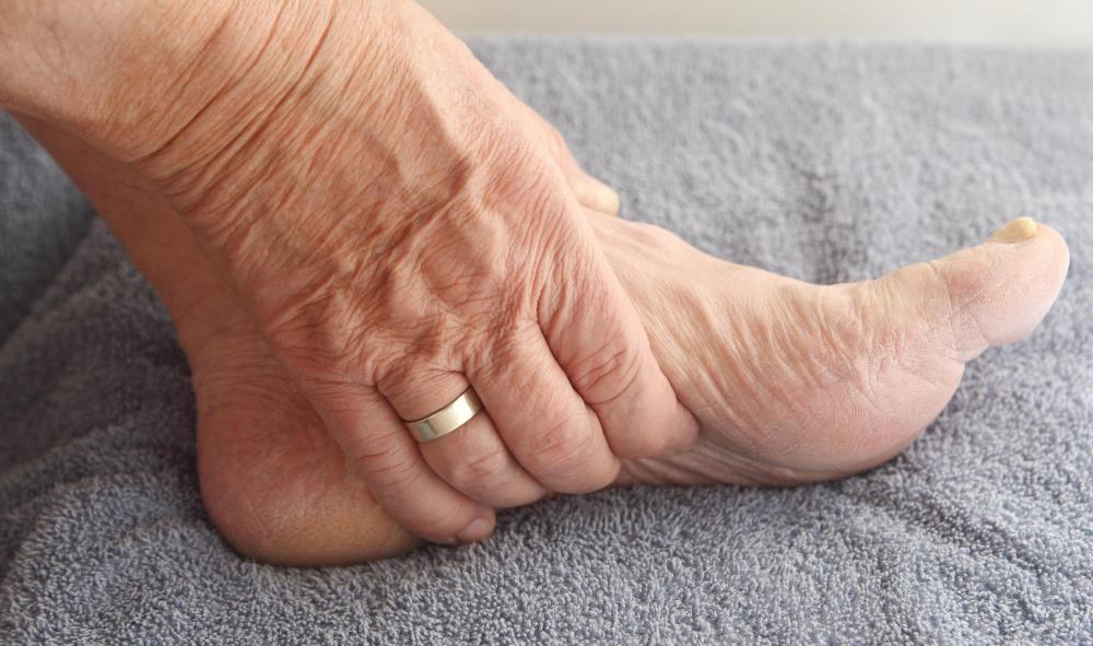
The veins of the leg contain small bicuspid valves that allow blood to flow in only one direction. This is analogous to a check valve in plumbing. The valve is closed by fluid that is flowing the wrong way. If these valves are damaged, small areas of turbulence can form, often leading to problems such as varicose veins.
AS FEATURED ON:
AS FEATURED ON:

-
![Arteries in the leg carry oxygenated blood, while veins carry deoxygenated blood.]() By: stockshoppeArteries in the leg carry oxygenated blood, while veins carry deoxygenated blood.
By: stockshoppeArteries in the leg carry oxygenated blood, while veins carry deoxygenated blood. -
![The inferior vena cava carries blood up to the heart.]() By: 7activestudioThe inferior vena cava carries blood up to the heart.
By: 7activestudioThe inferior vena cava carries blood up to the heart. -
![The veins of the legs can be divided into groups based on their functions and locations.]() By: Dmitry LobanovThe veins of the legs can be divided into groups based on their functions and locations.
By: Dmitry LobanovThe veins of the legs can be divided into groups based on their functions and locations. -
![The main veins of the leg include the femoral vein, the iliac veins, the popliteal vein, the tibial veins and the posterior arch vein.]() By: stockshoppeThe main veins of the leg include the femoral vein, the iliac veins, the popliteal vein, the tibial veins and the posterior arch vein.
By: stockshoppeThe main veins of the leg include the femoral vein, the iliac veins, the popliteal vein, the tibial veins and the posterior arch vein. -
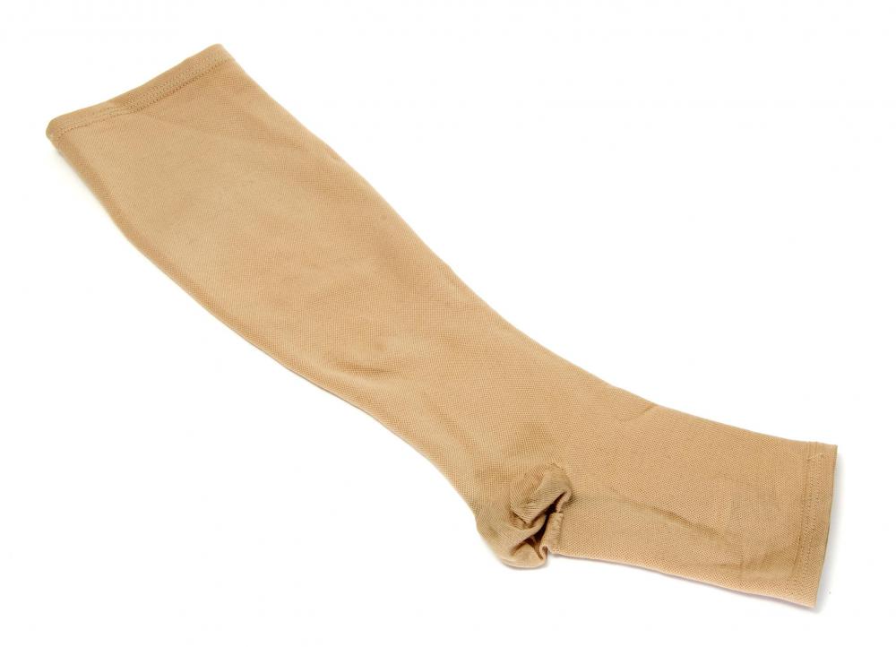
![Compression stockings may help relieve symptoms and slow progression of varicose veins.]() By: nitoCompression stockings may help relieve symptoms and slow progression of varicose veins.
By: nitoCompression stockings may help relieve symptoms and slow progression of varicose veins. -
![The saphenous veins meet in the dorsal venous foot arch.]() By: nebariThe saphenous veins meet in the dorsal venous foot arch.
By: nebariThe saphenous veins meet in the dorsal venous foot arch.


Discuss this Article
Post your comments