At WiseGEEK, we're committed to delivering accurate, trustworthy information. Our expert-authored content is rigorously fact-checked and sourced from credible authorities. Discover how we uphold the highest standards in providing you with reliable knowledge.
What are the Different Types of Dental Imaging?
In general, most dental images are created with X-ray technology, though there are several different types and formats within this broad category. The majority are created through what’s known as “intraoral” formatting, which is when the film is actually placed inside the mouth. “Extraoral” film is sometimes also used, though, and this is shot entirely externally. Dentists may use computed tomography (CT) imaging technology, too. These sorts of scans generate multiple images from different angles. The choice between the different types of images depends on a couple of different things, particularly the part of the mouth being scanned and the reason for taking the pictures in the first place. Simply creating images as a baseline for care is usually different than investigating known problems or conditions. Patient preferences, age, or other specifications might also factor in.
Imaging Basics

Dental imaging allows a dentist to be proactive in dental healthcare by seeing potential problems before they become visible or painful for the patient. In most cases, both the patient and the dentist benefit from catching a potential problem in the early stages. Imaging technology is non-invasive and patients don’t have to do anything special to prepare for it. Most dental offices have various imaging machines and devices on hand and scans are often done as part of routine check-up visits, though more complex or difficult diagnoses sometimes require visits to special imaging labs.
Intraoral X-Rays

The most common X-rays used in dental imaging are either intraoral or extraoral, depending on whether the X-ray film is inside or outside the mouth. These are typically further classified by what they show as either bitewing, periapical, or occlusal. Most of this classification centers on how the film is shot and the angle of the rays.
An intraoral X-ray, which is taken with the film inside the mouth, is usually the most common. This type of X-ray enables dentists and other trained technicians to see the details of the tooth, root and jaw area. It can also be used to view teeth that are developing, as in children.

Bitewing intraoral images usually focus on the crowns of the teeth in one section of the mouth and are typically used to check for tooth decay, bone density decay, and gum disease. Patients actually bite a small wing-shaped device to help channel the imaging rays in this sort of procedure. A periapical X-ray, by contrast, shows the entire teeth in one section, on either the top or the bottom of the mouth, and it can quickly make any root or bone structure problems pretty obvious. The third type of intraoral image is the occlusal view, which shows entire tooth development and placement.
Extraoral X-Rays

An extraoral X-ray is taken from the outside of the mouth. This type of scan does not have the detail of the intraoral X-ray but is very useful to find potential problems with the teeth and jaw. Extraoral scans can also show detail on impacted teeth and can highlight the relationship between the jaw and the teeth and can highlight potential problems that might exist between the two. One type of extraoral dental imaging X-ray is a panoramic shot, which shows the entire mouth. A tomogram scan, by contrast, shows only a particular portion in detail.
Special Projections

It is sometimes helpful for the dentist to see the patient’s entire head before making a diagnosis. A cephalometric projection is often the best choice in these situations. Orthodontists in particular often use this type of X-ray to work out comprehensive treatment plans for patients.
For some patients, a dentist will order a sialography X-ray to look for gland problems. This type of imaging requires the use of a dye, called a radiopaque contrast agent. The dye allows the soft tissue of the glands to show up on the film.
CT Scans
The last major tool a dentist uses for diagnostic purposes is the CT scan. This type of scan allows the inside of the head to be seen in a three-dimensional image. Unlike X-rays, though, these are normally conducted in a hospital setting instead of a dentist’s office, and tend to be much more expensive. CT scans are usually reserved for serious situations where getting a precise, multidimensional picture of the inner workings of the jaw and mouth is important to diagnosis and treatment.
AS FEATURED ON:
AS FEATURED ON:

-
![A dental X-ray machine.]() By: Konstantin ShevtsovA dental X-ray machine.
By: Konstantin ShevtsovA dental X-ray machine. -
![Dental x-rays are a common part of any dental examination.]() By: Volker WittDental x-rays are a common part of any dental examination.
By: Volker WittDental x-rays are a common part of any dental examination. -
![Dental x-rays help dentists diagnose teeth problems.]() By: belahocheDental x-rays help dentists diagnose teeth problems.
By: belahocheDental x-rays help dentists diagnose teeth problems. -
![Dental imaging can help with both diagnosis and prevention of future issues.]() By: Robert KneschkeDental imaging can help with both diagnosis and prevention of future issues.
By: Robert KneschkeDental imaging can help with both diagnosis and prevention of future issues. -
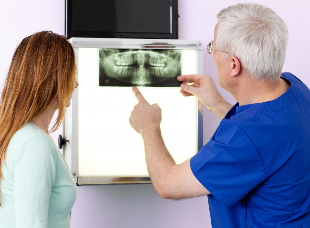
![A panoramic dental x-ray allows dentists to get a full view of all a patient's teeth.]() By: Georgiy PashinA panoramic dental x-ray allows dentists to get a full view of all a patient's teeth.
By: Georgiy PashinA panoramic dental x-ray allows dentists to get a full view of all a patient's teeth.


Discussion Comments
@spotiche5- Dental imaging is safe, and does not expose patients to high amounts of radiation. Today, the dental X-ray equipment that dentists use is more advanced than it use to be.
Whether you need bitewings or scans, you shouldn't hesitate get them. If you don't, you may put your dental health at risk because you may have cavities and other issues that need to be diagnosed and treated.
I haven't had dental imaging for several years because I am concerned about radiation levels. I know I need to have X-rays though because my dentist has recommended them. Does anyone have thoughts about the radiation levels caused by dental X-rays?
Post your comments