At TheHealthBoard, we're committed to delivering accurate, trustworthy information. Our expert-authored content is rigorously fact-checked and sourced from credible authorities. Discover how we uphold the highest standards in providing you with reliable knowledge.
What are Pain Receptors?
A pain receptor is a type of nerve cell that is primarily responsible for receiving and then transmitting stimulation signals from various nerve endings to the brain, which will typically interpret then as pain. Receptors work by releasing chemicals called “neurotransmitters” that course through the nerves, spinal column, and brain at very high speeds. The entire process of pain transmission is called nociception, and the pain receptors found in most bodily tissues are called nociceptors. Receptors are present in most parts of the body and respond to a wide range of stimuli; they are prolific in humans as well as most animals, and are the primary means through which pain is experienced.
Where They’re Found

Human beings have an extensive nervous system, which forms the basis for most sensations and many muscle movement and coordination tasks, too. Pain is a sensation that is usually experienced in association with some sort of damage or trauma and typically comes about as a result of nerve signals and chemical relays that begin with pain-specific receptors. These exist on almost all nerve endings though they tend to be most prolific in the skin, the muscles, and in joints; they’re also common in connective tissues and internal organs.

In most cases they are made up of only a few highly particularized cells, which makes them difficult to notice or observe without special equipment. Researchers know they exist, however, because of the dynamic ways in which they respond to stimulation and the steps through which they relay signals from even the farthest reaches of the body back to the pain center in the brain. On an individual level receptors are very small, then, but each is part of a large and dynamic system of signals and exchanges.
How They’re Activated

Receptors are activated in response to some sort of stimulation, whether internal or external. Pricking a finger with a pin is an example of external stimulation, while something like a twisted intestine or a blocked bowel is internal. The receptors on the ends of the nerves nearest where this stimulation takes place are usually the ones responsible for cataloging it and then sending it up the nerve into the main nervous system and, ultimately, to the brain.

When tissues or other body parts are damaged, they generally give off chemical substances known as “second messengers.” Important second messengers include bradykinin, prostaglandins, histamine, serotonin, leukotrienes, and potassium. When nerve endings sense the presence of these in the vicinity they typically activate their pain centers.
Differences in Nerve Type

The science behind how this all plays out can be somewhat complicated, but it often makes the most sense when taken on a nerve-by-nerve basis. The peripheral nerve fibers that contain these sorts of receptors are afferent nerves. This means that they send nerve impulses towards the brain and spinal cord. There are two main types of afferent nociceptors in the tissues: A-delta and C-sensory fibers.
The A-delta fibers are myelinated nerves, which means that they are covered by a slick protective shield; as a result these nerves generally transmit pain impulses very quickly. Pain receptors on A-delta fibers are activated in response to sharp, well-localized pain that requires an immediate reaction. This type of painful stimulus is sometimes referred to as “somatic pain,” and it usually involves damage to skin or muscle.
By contrast, the C-sensory pain fibers have receptors that are activated in response to dull, aching, or poorly localized pain stimuli. These pain fibers are unmyelinated, and as a result nerve impulses are generally transmitted more slowly. C-sensory nerve fibers respond to so-called “visceral pain,” which is usually caused by damage to internal organs.
Pain Signal Journey
Once the second messenger stimulus is transmitted along afferent nerves, it must go through the dorsal horn of the spinal cord. This is called the “relay station” for pain signals and is where the painful stimuli are transmitted to different parts of the brain. Some pain impulses are transmitted directly to the thalamus and brain stem for a quick response, while others are sent to the frontal cortex of the brain for further processing. It is in the frontal cortex that the conscious realization of pain takes place.
The Brain’s Response
The final step in the pain transmission process is a response from the brain to tell the body how to react. These instructions are carried as impulses along efferent nerves, away from the brain. During pain transmission, many substances may be released in the brain and spinal cord that either increase or decrease the perception of the painful stimulus. These are called neurochemical mediators, and include endorphins, which are natural analgesics, as well as serotonin and norepinephrine, which enhance a person’s perception of pain.
Pain Killer Basics
Pain medications, sometimes called “pain killers,” usually work by targeting the secondary messengers and neurochemical mediators. If a drug inhibits the release of second messengers, then the pain receptors will not be activated, the pain impulse will not reach the brain, and the person will not perceive pain from the damaged tissue. The same thing happens if the brain’s response is delayed or neutralized. In most cases medications can only offer temporary fixes, and aren’t usually able to cure the underlying problem. All they do is prevent a person from feeling the pain associated with the injury or damage.
AS FEATURED ON:
AS FEATURED ON:

-
![Muscles are one type of tissue with pain receptors.]() By: MaridavMuscles are one type of tissue with pain receptors.
By: MaridavMuscles are one type of tissue with pain receptors. -
![Pain receptors in the stomach are activated in response to a stimulus.]() By: Ana Blazic PavlovicPain receptors in the stomach are activated in response to a stimulus.
By: Ana Blazic PavlovicPain receptors in the stomach are activated in response to a stimulus. -
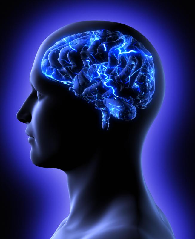
![Pain receptors send signals to the brain.]() By: James SteidlPain receptors send signals to the brain.
By: James SteidlPain receptors send signals to the brain. -
![Humans have extensive, complex nervous systems that form the basis for pain and other sensations.]() By: AllianceHumans have extensive, complex nervous systems that form the basis for pain and other sensations.
By: AllianceHumans have extensive, complex nervous systems that form the basis for pain and other sensations. -
![Pain receptors are most prolific in the skin.]() By: Deyan GeorgievPain receptors are most prolific in the skin.
By: Deyan GeorgievPain receptors are most prolific in the skin.


Discussion Comments
Simple and comprehensive.
who wrote this? just wondering. i need it for my school Interest Project.
Very interesting article!
Helpful. Thanks.
Post your comments