At WiseGEEK, we're committed to delivering accurate, trustworthy information. Our expert-authored content is rigorously fact-checked and sourced from credible authorities. Discover how we uphold the highest standards in providing you with reliable knowledge.
How does MRI Scanning Work?
 Jessica Ellis
Jessica Ellis
Jessica Ellis
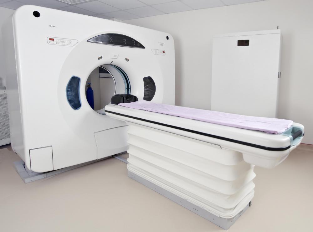
Magnetic Resonance Imagery (MRI) scanning is an advanced medical technique that is used to produce high resolution images of the interior of the body. Unlike an X-ray, an MRI image can show the soft tissues of the body, while also having the flexibility to examine very small areas of the body from a wide variety of angles. MRI scanning works through the combination of enormous magnets, carefully targeted electromagnetic pulses, and computer software that turns raw data into finished images. Many medical experts credit MRI scanning with revolutionizing the diagnostic field of medicine.
It may not feel like it, but every person is comprised of billions of atoms, all working busily to create and maintain the physical body. Human beings are mostly comprised of water, which itself is made up of a combination of two hydrogen atoms and one oxygen atom. Hydrogen atoms, of which the body has many, spin randomly under normal circumstances. When subjected to an attuned magnet, however, most of the hydrogen atoms will stop their random meanderings and point to the same position, aligning with the direction of the magnetic field. The first step of MRI scanning is to create a magnetic field that aligns the hydrogen atoms, usually making about half point toward the feet, and half toward the head.

MRI scanning relies on the fact that a very few hydrogen atoms will refuse to line up with their billions of atomic brethren. These few continue to spin randomly after the magnetic field is applied, making them stand out from the pack. Using a radio frequency pulse, the MRI machine targets the still-random atoms, which absorb the energy of the pulse and spin in a different direction. An array of smaller magnets in the machine, known as gradients, spring to life during this process, localizing the efforts of the machine on the specific part of the body that needs to be examined.

The final step in MRI scanning is the creation of the image. After the gradients have focused in on the slice of the body that needs attention, the radio pulses are stopped, allowing the atoms to expel the energy they have absorbed and rotate back to their original position. The machine measures several different variables of their rate of return to the original equilibrium, and it is these measurements that provide the raw data to create the final image.

The final image is a product of computer wizardry and medical technology. Patients are often injected with a contrast agent that stains different types of tissue different shades, so that contrasts will show up on the created image. Depending on the computer system used, the information garnered from the MRI scanning can be turned into a two or three dimensional image, which illuminates tissue distinctions thanks to the contrast agent.

Though MRI scanning is considered a very safe procedure that often produces excellent results, there are some drawbacks to the process. First, the scanning requires that the patient lie perfectly still, or else the image will be disrupted. While this may not seem like a large requirement, it is often made more difficult by the fact that the machine is very loud and places the patient in a small, enclosed space. People uncomfortable with tight spaces may want to ask doctors about possible options to ease the process.
Jessica Ellis
With a B.A. in theater from UCLA and a graduate degree in screenwriting from the American Film Institute, Jessica is passionate about drama and film. She has many other interests, and enjoys learning and writing about a wide range of topics in her role as a WiseGEEK writer.
Learn more...
Jessica Ellis
With a B.A. in theater from UCLA and a graduate degree in screenwriting from the American Film Institute, Jessica is passionate about drama and film. She has many other interests, and enjoys learning and writing about a wide range of topics in her role as a WiseGEEK writer.
Learn more...AS FEATURED ON:
AS FEATURED ON:

-
![A medical professional reviews MRI scans for signs of internal injuries.]() By: Konstantin SutyaginA medical professional reviews MRI scans for signs of internal injuries.
By: Konstantin SutyaginA medical professional reviews MRI scans for signs of internal injuries. -
![A MRI scan of the brain.]() By: Marcin SadlowskiA MRI scan of the brain.
By: Marcin SadlowskiA MRI scan of the brain. -
![An MRI machine.]() By: Hakan KızıltanAn MRI machine.
By: Hakan KızıltanAn MRI machine. -
![The radiologist or technician can view the MRI images in real time from the observation room.]() By: EPSTOCKThe radiologist or technician can view the MRI images in real time from the observation room.
By: EPSTOCKThe radiologist or technician can view the MRI images in real time from the observation room.


Discuss this Article
Post your comments