At TheHealthBoard, we're committed to delivering accurate, trustworthy information. Our expert-authored content is rigorously fact-checked and sourced from credible authorities. Discover how we uphold the highest standards in providing you with reliable knowledge.
How do I Interpret my PET Scan Results?
A positron emission tomography scan, also known as a PET scan, is a non-invasive imaging test that uses a radioactive tracer to reveal various diseases in the body. PET scan results are generally only interpreted by radiologists who have received specialized training in nuclear medicine — a type of medicine that uses small amounts of radioactive substances, called radiotracers or radiopharmaceuticals. After such analysis, radiologists usually forward the PET scan results to the ordering physician, who then typically reviews them with the patient.
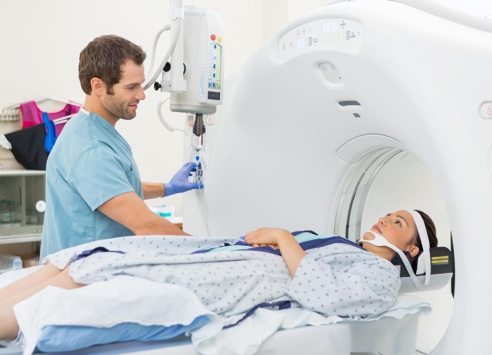
In order to prepare for a PET scan, a patient must first receive a radiotracer. Depending on a variety of circumstances, this can either be given to the patient intravenously, as an inhaled gas, or as a drinkable liquid. Once the radiotracer settles in the area to be imaged, it emits a certain type of energy known as a gamma ray. A gamma camera, PET scanner, and probe measure the gamma rays. A computer then helps to determine how much radiotracer is being absorbed by those areas. The computer also takes pictures of the targeted areas, which indicates its structure and functioning ability, including blood flow, oxygen usage, and glucose metabolism.

When a radiologist interprets PET scan results, he or she may be able to identify several different diseases, including cancer, as the test typically highlights the most rapidly growing cells in the body. These conditions light up as hot spots on the images that the radiologist can see on a computer. The activity of such spots is usually expressed in Standardized Uptake Value (SUV), which help the radiologist interpret the scan results. Generally speaking, cancer will have an SUV of over 2.5, though there are a variety of other factors that may influence how a radiologist interprets any given hot spot. These factors can include the location of the suspected cancer, the patient's history, and other imaging results.

PET scan images can be combined with computed tomography (CT) images or magnetic resonance imaging (MRI) in a process called co-registration or image fusion. Most PET scans are currently combined with CT scans to create images that provide detailed anatomical information about the organs and tissues. Results from PET/CT scans typically give more accurate diagnostic and treatment information than PET scan results alone. PET and PET/CT scans are most commonly used to help diagnose and re-stage cancer, evaluate the heart muscle, and detect brain abnormalities. In all cases, interpreting PET/CT or PET scan results can be a very complicated process and should generally be reserved for physicians and radiologists who have received specialized training in nuclear medicine.
AS FEATURED ON:
AS FEATURED ON:

-
![PET scans should be interpreted by an experienced physician.]() By: griezePET scans should be interpreted by an experienced physician.
By: griezePET scans should be interpreted by an experienced physician. -
![Most PET scans are currently combined with CT scans.]() By: Konstantin SutyaginMost PET scans are currently combined with CT scans.
By: Konstantin SutyaginMost PET scans are currently combined with CT scans. -
![In most cases PET scan results will not be available for several days, although in emergency situations they may be interpreted in real time.]() By: EPSTOCKIn most cases PET scan results will not be available for several days, although in emergency situations they may be interpreted in real time.
By: EPSTOCKIn most cases PET scan results will not be available for several days, although in emergency situations they may be interpreted in real time. -
![CT scans are often used with PET scans to diagnose cancer, examine heart muscle and detect brain abnormalities.]() By: Tyler OlsonCT scans are often used with PET scans to diagnose cancer, examine heart muscle and detect brain abnormalities.
By: Tyler OlsonCT scans are often used with PET scans to diagnose cancer, examine heart muscle and detect brain abnormalities. -
![An MRI can be used in conjunction with a PET scan to get an accurate picture of the brain.]() By: Mikhail BasovAn MRI can be used in conjunction with a PET scan to get an accurate picture of the brain.
By: Mikhail BasovAn MRI can be used in conjunction with a PET scan to get an accurate picture of the brain. -
![PET scan results are generally only interpreted by radiologists who have received specialized training in nuclear medicine.]() By: michaeljungPET scan results are generally only interpreted by radiologists who have received specialized training in nuclear medicine.
By: michaeljungPET scan results are generally only interpreted by radiologists who have received specialized training in nuclear medicine. -
![A PET scanner uses gamma cameras to capture images inside the body.]() By: sonapA PET scanner uses gamma cameras to capture images inside the body.
By: sonapA PET scanner uses gamma cameras to capture images inside the body.


Discussion Comments
What does this mean?
1. Small focal area of cutaneous hypermetabolic activity over the left maxilla with maximal SUV of 3.7. Given history of melanoma, further evaluation of this site is recommended.
2. Small focus of hypermetabolic activity in the left fourth rib, max motion be of 2.8. Given the lack of a visible abnormality on the CT map the significance is uncertain. Consider correlation with bone scan SPECT versus follow-up PET scan.
What does a 1.2 (total body) and 1.5 (chest) number mean? Seems very low. They can't do a needle biopsy or other procedure other than a complete (right lower lobe) lobectomy, to get a diagnosis, for at least 9 months (post stent anticoagulants). I refused and said let's repeat this later. (smoker, lung CT with 1.0 cm nodule found incidentally). The lesion had central lucency.
I had a chest CT scan "because I'm a smoker." Out of the blue, it showed a 1.5 cm x 1.0 cm nodule in the RLL. The PET scan came back 1.2 and 1.5. It showed a central (quiet) lucent area. I had just had stents and am on an anticoagulant for one year, at least. I cannot have a needle biopsy or endobronchial biopsy because cessation of the anticoagulant has too big a risk for death at this time. The pulmonologist was all go for a right lower lobectomy. Just, if for no other reason, to get a diagnosis (a wedge biopsy or sementectomy are also contraindicated in someone on anticoagulants). I do understand the reasoning here. But I am reluctant to forsake 30 percent of my pulmonary function just to find out I aspirated a garbonzo bean.
So, does anybody have any unbiased opinion on what a 1.2 (body) 1.5 (chest) uptake may indicate?
What does it mean if your doctor tells you your pet scan numbers have gone from a 3.5 to a 4? I don't understand.
Breast cancer runs in my family and I've actually seen quite a few PET and CT scanning results until now. But I only know two acronym that is used in PET scans and that is "NED" and "Progress." NED is a lovely acronym to see in PET scans because it means "No Evidence of Disease." This is what every cancer patient (or anyone having a PET scan) wants to see on their PET scan results!
"Progress" sounds like it's a positive thing, but it's not. It stands for "progression of metastases" which means that the cancer is spreading.
My sister said that it's possible to get a list of the acronyms used in PET scans from various resources but I've not looked into it.
@simrin-- I know what you mean. I'm a lung cancer survivor and I remember the last PET lung scan results I received from the lab. I had no idea what it was talking about, it was like a foreign language! And all my doctor said was that it was looking good and I would be off the medications.
The person who actually diagnosis diseases is the radiologist. They're the ones who can tell what's what. The physicians receive the diagnosis from them and simply assist patients with treatment, which they often cooperate with other doctors and experts to do.
Most people never even see their radiologist but if you're not getting much information from your physician, you can request to see your radiologist. I saw my radiologist twice and he was amazing. He described everything to me in simple and clear terms and I felt a lot better after speaking to him.
I have several friends and family members who were very confused when they got their PET scan results because their physician didn't explain much to them. For some reason, physicians don't often go into the details of the results but rather just tell the patient what the next steps will be.
If there isn't much of concern, they'll say, "you're fine" or "come back in 3 months for a check-up." If there is something more serious, then they'll somewhat explain the situation and recommend treatment options.
But this not always enough and I think people deserve to know the details of their situation, especially if it's something like a cancer related PET scan. And it's not like people can analyze PET scan results on their own! Even the physicians can't do it! Only the radiologist can!
Post your comments